
TNFRSF10A Antibody, Unconjugated, Rabbit, Polyclonal
Catalog Number:
BYT-ORB1239340
- Images (11)


| Article Name: | TNFRSF10A Antibody, Unconjugated, Rabbit, Polyclonal |
| Biozol Catalog Number: | BYT-ORB1239340 |
| Supplier Catalog Number: | orb1239340 |
| Alternative Catalog Number: | BYT-ORB1239340-0.02,BYT-ORB1239340-0.1 |
| Manufacturer: | Biorbyt |
| Host: | Rabbit |
| Category: | Antikörper |
| Application: | ELISA, ICC, IF, IHC-P, WB |
| Species Reactivity: | Human, Mouse, Rat |
| Immunogen: | Anti-DR4 antibody (orb1239340) was raised against a peptide corresponding to 19 amino acids near the carboxy terminus of human DR4. The immunogen is located within the last 50 amino acids of DR4. |
| Conjugation: | Unconjugated |
| Alternative Names: | DR4 Antibody: DR4, APO2, CD261, TRAILR1, TRAILR-1, DR4, Tumor necrosis factor receptor superfamily member 10A, Death receptor 4, TRAIL receptor 1 |
| TNFRSF10A Antibody |
|
|
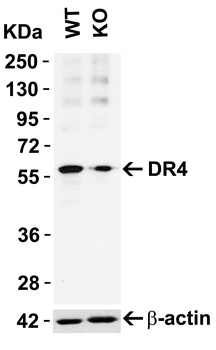
KO Validation in HeLa Cells. Loading: 10 µg of HeLa WT cell lysates or DR4 KO cell lysates. Antibodies: DR4 orb1239340 (1 µg/mL) and beta-actin orb1240312 (1 µg/mL), 1 h incubation at RT in 5% NFDM/TBST. Secondary: Goat Anti-Rabbit IgG HRP conjugate at 1:10000 dilution. |
|
|
KD Validation in Huh7 cells (Malhi et al., 2007). Western blot analysis with anti-DR4 antibodies (orb1239340) was performed for DR5 and DR4 expression using whole cell lysates from Huh 7 cells transfected with respective siRNAs. In cells treated with siDR4, a decrease in DR4 level was observed, DR5 levels were unchanged. Scrambled siRNA was used as control. |
|
|
KD Validation in HeLa cells (Horinaka et al., 2005). HeLa cells were transfected with DR4siRNA or LacZ control siRNA. At 24 h after transfection, the cells were treated with or without 20 µM luteolin for 24 h. Western blot analysis was carried out with anti-DR4 antibodies (orb1239340). DR4 expression was markedly reduced after DR4 knockdown. |
|
|
Independent Antibody Validation (IAV) via Protein Expression Profile in Cell Lines. Loading: 15 µg of lysates per lane. Antibodies: DR4 orb1239340 (1 µg/mL), DR4 orb1239343 (4 µg/mL), and beta-actin (1 µg/mL), 1h incubation at RT in 5% NFDM/TBST. Secondary: Goat anti-rabbit IgG HRP conjugate at 1:10000 dilution. |
|
|
Western Blot Validation in Cell Lines. Loading: 15 µg of cell lysates per lane. Antibodies: DR4 orb1239340 (1 µg/mL), 1h incubation at RT in 5% NFDM/TBST. Secondary: Goat anti-rabbit IgG HRP conjugate at 1:10000 dilution. |
|
|
Immunofluorescence Validation of DR4. Immunofluorescent analysis of 4% paraformaldehyde-fixed human spleen tissue labeling DR4 with orb1239340 at 20 µg/mL, followed by goat anti-rabbit IgG secondary antibody at 1/500 dilution (red) and DAPI staining (blue). Image showing membrane staining on human spleen cells. |
|
|
Immunocytochemistry Validation of DR4 in HeLa Cells. Immunocytochemical analysis of HeLa cells using anti-DR4 antibody (orb1239340) at 10 µg/ml. Cells was fixed with formaldehyde and blocked with 10% serum for 1 h at RT, antigen retrieval was by heat mediation with a citrate buffer (pH6). Samples were incubated with primary antibody overnight at 4C. A goat anti-rabbit IgG H&L (HRP) at 1/250 was used as secondary. Counter stained with Hematoxylin. |
|
|
Immunohistochemistry Validation of DR4. Immunohistochemical analysis of paraffin-embedded human spleen tissue using anti-DR4 antibody (orb1239340) at 10 µg/ml. Tissue was fixed with formaldehyde and blocked with 10% serum for 1 h at RT, antigen retrieval was by heat mediation with a citrate buffer (pH6). Samples were incubated with primary antibody overnight at 4C. A goat anti-rabbit IgG H&L (HRP) at 1/250 was used as secondary. Counter stained with Hematoxylin. |
|
|
KD Validation in SW480 cells (Goda et al., 2008). The expression of DR4 was knocked down via DR4 siRNA, 24 h latercells were treated with dipyridamole for 24 h. DR4 protein expression detected by anti-DR4 antibodies (orb1239340) was disrupted. Dipyridamole up-regulated the expression of DR4. |
|
|
Immunofluorescence Validation of DR4 in rat brain (Cantarella et al., 2014). DR4 protein expression detected by anti-DR4 antibodies (orb1239340) was increased after transient brain ischemia (tMCAO) and decreased after pre-conditioning stimulus. Confocal microscopic images displaying NeuN (a, d, g) (green), DR4 (b, e, h) (red), and Merge (c, f, i) (yellow) in the brain peri-ischemic region of rats after 5 h. |
|
|
Immunocytochemistry Validation of DR4 in human melanoma cells (Ekmekcioglu et al., 2008). MeWo melanoma cells were exposed to affinity-purified MDA7/IL-24. After 48 h of treatment, cells were collected and cytospins prepared for cytochemical assessment of their TRAIL receptor (R1 and R2) expression (anti-DR4 (orb1239340) or anti-DR5, AEC, hematoxylin). Both DR4 and DR5 expression were upregulated in MeWo cells after treatment. |
Product Guarantee and Expert Support
