Nhe-1 antibody was raised against a 17 amino acid synthetic peptide near the center of the human Nhe-1.The immunogen is located within amino acids 490 - 540 of Nhe-1.
Nhe-1 Antibody is supplied in PBS containing 0.02% sodium azide.
Form:
Liquid
Target:
SLC9A1
Application Notes:
Application Notes: Nhe-1 antibody can be used for detection of Nhe-1 by Western blot at 1 - 2 µg/mL. Antibody can also be used for immunohistochemistry starting at 2.5 µg/mL. For immunofluorescence start at 20 µg/mL.Antibody validated: Western Blot in human samples, Immunohistochemistry in human samples and Immunofluorescence in human samples. All other applications and species not yet tested
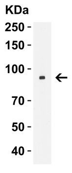
WB Validation in K562 Cells. Loading: 10 µg of lysate. Antibodies: Nhe-1, orb1239375, 4 µg/mL, 1 h incubation at RT in 5% NFDM/TBST. Secondary: Goat Anti-Rabbit IgG HRP conjugate at 1:10000 dilution.
Immunohistochemical staining of human brain tissue using Nhe-1 antibody at 2.5 µg/mL.
WB Validation in Mouse Tissues. Loading: 15 µg of lysate. Antibodies: Nhe-1, orb1239375, 1 µg/mL, 1 h incubation at RT in 5% NFDM/TBST. Secondary: Goat Anti-Rabbit IgG HRP conjugate at 1:10000 dilution.
WB Validation in Rat Brain. Loading: 15 µg of lysate. Antibodies: Nhe-1, orb1239375, 1 µg/mL, 1 h incubation at RT in 5% NFDM/TBST. Secondary: Goat Anti-Rabbit IgG HRP conjugate at 1:10000 dilution.
Immunohistochemistry Validation of Nhe-1 in Human Small Intestine. Immunohistochemical analysis of paraffin-embedded human small intestine tissue using anti-Nhe-1 antibody (orb1239375) at 2 µg/ml. Tissue was fixed with formaldehyde and blocked with 10% serum for 1 h at RT, antigen retrieval was by heat mediation with a citrate buffer (pH6). Samples were incubated with primary antibody overnight at 4C. A goat anti-rabbit IgG H&L (HRP) at 1/250 was used as secondary. Counter stained with Hematoxylin.
Immunohistochemistry Validation of Nhe-1 in Mouse Colon. Immunohistochemical analysis of paraffin-embedded mouse colon tissue using anti-Nhe-1 antibody (orb1239375) at 1 µg/ml. Tissue was fixed with formaldehyde and blocked with 10% serum for 1 h at RT, antigen retrieval was by heat mediation with a citrate buffer (pH6). Samples were incubated with primary antibody overnight at 4C. A goat anti-rabbit IgG H&L (HRP) at 1/250 was used as secondary. Counter stained with Hematoxylin.
Immunohistochemistry Validation of Nhe-1 in Rat Small Intestine. Immunohistochemical analysis of paraffin-embedded rat small intestine tissue using anti-Nhe-1 antibody (orb1239375) at 1 µg/ml. Tissue was fixed with formaldehyde and blocked with 10% serum for 1 h at RT, antigen retrieval was by heat mediation with a citrate buffer (pH6). Samples were incubated with primary antibody overnight at 4C. A goat anti-rabbit IgG H&L (HRP) at 1/250 was used as secondary. Counter stained with Hematoxylin.
* VAT and and shipping costs not included. Errors and price changes excepted