GLS2 antibody was raised against an 18 amino acid synthetic peptide near the center terminus of human GLS2.The immunogen is located within amino acids 300 - 350 of GLS2.
GLS2 Antibody is supplied in PBS containing 0.02% sodium azide.
Form:
Liquid
Target:
GLS2
Application Notes:
Application Notes: GLS2 antibody can be used for detection of GLS2 by Western blot at 0.5 - 1 µg/mL. Antibody can also be used for immunohistochemistry starting at 5 µg/mL. For immunofluorescence start at 20 µg/mL.Antibody validated: Western Blot in rat samples, Immunohistochemistry in mouse and rat samples and Immunofluorescence in mouse and rat samples. All other applications and species not yet tested
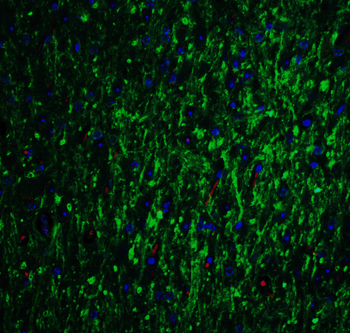
Immunofluorescence Validation of GLS2 in Mouse Brain. Immunofluorescent analysis of 4% paraformaldehyde-fixed mouse brain labeling GLS2 with orb1239416 at 20 µg g/mL, followed by goat anti-rabbit IgG secondary antibody at 1/500 dilution (green) and DAPI antibody (blue).
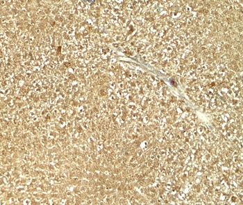
Immunohistochemistry of GLS2 in rat liver tissue with GLS2 antibody at 5 µg/mL.
Immunohistochemistry Validation of GLS2 in Human Liver. Immunohistochemical analysis of paraffin-embedded human liver tissue using anti-GLS2 antibody (orb1239416) at 1 µg/mL. Tissue was fixed with formaldehyde and blocked with 10% serum for 1 h at RT, antigen retrieval was by heat mediation with a citrate buffer (pH6). Samples were incubated with primary antibody overnight at 4C. A goat anti-rabbit IgG H&L (HRP) at 1/250 was used as secondary. Counter stained with Hematoxylin.
Immunohistochemistry Validation of GLS2 in Mouse Liver. Immunohistochemical analysis of paraffin-embedded mouse liver tissue using anti-GLS2 antibody (orb1239416) at 1 µg/mL. Tissue was fixed with formaldehyde and blocked with 10% serum for 1 h at RT, antigen retrieval was by heat mediation with a citrate buffer (pH6). Samples were incubated with primary antibody overnight at 4C. A goat anti-rabbit IgG H&L (HRP) at 1/250 was used as secondary. Counter stained with Hematoxylin.
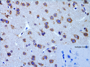
Immunohistochemistry Validation of GLS2 in Rat Brain. Immunohistochemical analysis of paraffin-embedded rat brain tissue using anti-GLS2 antibody (orb1239416) at 2 µg/mL. Tissue was fixed with formaldehyde and blocked with 10% serum for 1 h at RT, antigen retrieval was by heat mediation with a citrate buffer (pH6). Samples were incubated with primary antibody overnight at 4C. A goat anti-rabbit IgG H&L (HRP) at 1/250 was used as secondary. Counter stained with Hematoxylin.
WB Validation in Human Pancreas. Loading: 10 µg of lysate Antibodies: GLS2, orb1239416, 2 µg/mL, 1 h incubation at RT in 5% NFDM/TBST. Secondary: Goat Anti-Rabbit IgG HRP conjugate at 1:10000 dilution.
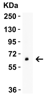
WB Validation in Mouse and Rat Brain. Loading: 15 µg of lysate Antibodies: GLS2, orb1239416, 1 µg/mL, 1 h incubation at RT in 5% NFDM/TBST. Secondary: Goat Anti-Rabbit IgG HRP conjugate at 1:10000 dilution.
* VAT and and shipping costs not included. Errors and price changes excepted