
ERN1 Antibody, Unconjugated, Rabbit, Polyclonal
Catalog Number:
BYT-ORB1239523
- Images (11)


| Article Name: | ERN1 Antibody, Unconjugated, Rabbit, Polyclonal |
| Biozol Catalog Number: | BYT-ORB1239523 |
| Supplier Catalog Number: | orb1239523 |
| Alternative Catalog Number: | BYT-ORB1239523-0.02,BYT-ORB1239523-0.1 |
| Manufacturer: | Biorbyt |
| Host: | Rabbit |
| Category: | Antikörper |
| Application: | ELISA, ICC, IF, IHC-P, WB |
| Species Reactivity: | Human, Mouse, Rat |
| Immunogen: | Anti-IRE1p antibody (orb1239523) was raised against a peptide corresponding to 16 amino acids near the carboxy terminus of human IRE1P. The immunogen is located within the last 50 amino acids of IRE1p. |
| Conjugation: | Unconjugated |
| Alternative Names: | IRE1p Antibody: IRE1, IRE1P, IRE1a, hIRE1p, IRE1, Endoplasmic reticulum-to-nucleus signaling 1 |
| ERN1 Antibody |
|
|
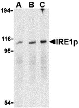
Western Blot Validation in Mouse A20 Cell Lysate. Loading: 15 µg of lysates per lane. Antibodies: IRE1p orb1239523 (A: 0.5 µg/mL, B: 1 µg/mL, C: 2 µg/mL), 1h incubation at RT in 5% NFDM/TBST. Secondary: Goat anti-rabbit IgG HRP conjugate at 1:10000 dilution. |
|
|
Immunohistochemistry Validation of IRE1p in Rat Small Intestine Tissue. Immunohistochemical analysis of paraffin-embedded Rat Small Intestine Tissue using anti-IRE1P antibody (orb1239523) at 2 µg/ml. Tissue was fixed with formaldehyde and blocked with 10% serum for 1 h at RT, antigen retrieval was by heat mediation with a citrate buffer (pH6). Samples were incubated with primary antibody overnight at 4C. A goat anti-rabbit IgG H&L (HRP) at 1/250 was used as secondary. Counter stained with Hematoxylin. |
|
|
Induced Expression Validation of IRE1p in human umbilical vein endothelial cells (HUVECs) (Wang et al., 2019). IRE1p expression was examined by Western blot analysis with anti-IRE1p antibodies (orb1239523). IRE1p was increased in HUVEC cells treated with 10 µM 20 (S)-PPD for 6 to 8 hours compared with control cells. |
|
|
KO Validation in HeLa Cells. Loading: 10 µg of WT cell lysates (lane 1) or IRE1P KO cell lysates (lane 2). Antibodies: IRE1P orb1239523 (0.5 µg/mL) and beta-actin (1 µg/mL), 1h incubation at RT in 5% NFDM/TBST. Secondary: Goat anti-rabbit IgG HRP conjugate at 1:10000 dilution. |
|
|
Western Blot Validation in Human Cell Lines. Loading: 15 µg of lysates per lane. Antibodies: IRE1p orb1239523 (0.4 µg/mL), 1h incubation at RT in 5% NFDM/TBST. Secondary: Goat anti-rabbit IgG HRP conjugate at 1:10000 dilution. Lane 1: Caco-2, Lane2: SK-N-SH. |
|
|
Western Blot Validation in Rat Brain Tissue Lysate. Loading: 15 µg of lysates per lane. Antibodies: IRE1p orb1239523 (A: 0.5 µg/mL, B: 1 µg/mL), 1h incubation at RT in 5% NFDM/TBST. Secondary: Goat anti-rabbit IgG HRP conjugate at 1:10000 dilution. |
|
|
Immunofluorescence Validation of IRE1p in Mouse A20 Cells. Immunofluorescent analysis of 4% paraformaldehyde-fixed A20 Cells labeling IRE1P with orb1239523 at 2 µg/mL, followed by goat anti-rabbit IgG secondary antibody at 1/500 dilution (red). |
|
|
Immunocytochemistry Validation of IRE1p in Mouse A20 Cells. Immunocytochemical analysis of A20 cells using anti-IRE1p antibody (orb1239523) at 1 µg/ml. Cells was fixed with formaldehyde and blocked with 10% serum for 1 h at RT, antigen retrieval was by heat mediation with a citrate buffer (pH6). Samples were incubated with primary antibody overnight at 4C. A goat anti-rabbit IgG H&L (HRP) at 1/250 was used as secondary. Counter stained with Hematoxylin. |
|
|
Immunofluorescence Validation of IRE1p in Human Small Intestine Tissue. Immunofluorescent analysis of 4% paraformaldehyde-fixed Human Small Intestine Tissue labeling IRE1p with orb1239523 at 20 µg/mL, followed by goat anti-rabbit IgG secondary antibody at 1/500 dilution (green) and DAPI staining (blue). |
|
|
Immunohistochemistry Validation of IRE1p in Human Small Intestine Tissue. Immunohistochemical analysis of paraffin-embedded Human Small Intestine Tissue using anti-IRE1P antibody (orb1239523) at 2 µg/ml. Tissue was fixed with formaldehyde and blocked with 10% serum for 1 h at RT, antigen retrieval was by heat mediation with a citrate buffer (pH6). Samples were incubated with primary antibody overnight at 4C. A goat anti-rabbit IgG H&L (HRP) at 1/250 was used as secondary. Counter stained with Hematoxylin. |
|
|
Immunofluorescence Validation of IRE1p in Rat Small Intestine Tissue. Immunofluorescent analysis of 4% paraformaldehyde-fixed Rat Small Intestine Tissue labeling IRE1p with orb1239523 at 20 µg/mL, followed by goat anti-rabbit IgG secondary antibody at 1/500 dilution (green) and DAPI staining (blue). |
Product Guarantee and Expert Support
