
IRF7 Antibody, Unconjugated, Rabbit, Polyclonal
Catalog Number:
BYT-ORB1239524
- Images (11)


| Article Name: | IRF7 Antibody, Unconjugated, Rabbit, Polyclonal |
| Biozol Catalog Number: | BYT-ORB1239524 |
| Supplier Catalog Number: | orb1239524 |
| Alternative Catalog Number: | BYT-ORB1239524-0.02,BYT-ORB1239524-0.1 |
| Manufacturer: | Biorbyt |
| Host: | Rabbit |
| Category: | Antikörper |
| Application: | ELISA, IHC-P, WB |
| Species Reactivity: | Human, Mouse, Rat |
| Immunogen: | Anti-IRF7 antibody (orb1239524) was raised against a peptide corresponding to 17 amino acids near the center of human IRF7. The immunogen is located within amino acids 200-230 of IRF7. |
| Conjugation: | Unconjugated |
| Alternative Names: | IRF7 Antibody: IRF7A, IRF7B, IRF7C, IRF7H, IRF-7H, Interferon regulatory factor 7, IRF-7 |
| IRF7 Antibody |
|
|
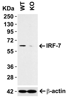
KO Validation in A549 Cell Lysate. Loading: 10 µg of A549 WT cell lysates or IRF7 KO cell lysates. Antibodies: IRF7 orb1239524 (0.5 µg/mL) and beta-actin orb1240312 (1 µg/mL), 1 h incubation at RT in 5% NFDM/TBST. Secondary: Goat Anti-Rabbit IgG HRP conjugate at 1:10000 dilution. |
|
|
Immunohistochemistry Validation of IRF7 in Mouse Pancreas Tissue. Immunohistochemical analysis of paraffin-embedded mouse pancreas tissue using anti-IRF7 antibody (orb1239524) at 2 µg/ml. Tissue was fixed with formaldehyde and blocked with 10% serum for 1 h at RT, antigen retrieval was by heat mediation with a citrate buffer (pH6). Samples were incubated with primary antibody overnight at 4 C. A goat anti-rabbit IgG H&L (HRP) at 1/250 was used as secondary. Counter stained with Hematoxylin. |
|
|
Immunohistochemistry Validation of IRF7 in Rat Brain Tissue. Immunohistochemical analysis of paraffin-embedded rat brain tissue using anti-IRF7 antibody (orb1239524) at 5 µg/ml. Tissue was fixed with formaldehyde and blocked with 10% serum for 1 h at RT, antigen retrieval was by heat mediation with a citrate buffer (pH6). Samples were incubated with primary antibody overnight at 4 C. A goat anti-rabbit IgG H&L (HRP) at 1/250 was used as secondary. Counter stained with Hematoxylin. |
|
|
Western Blot Validation in Human Cell Lines. Loading: 15 µg of lysates per lane. Antibodies: IRF7, orb1239524, (1 µg/mL), 1h incubation at RT in 5% NFDM/TBST. Secondary: Goat anti-rabbit IgG HRP conjugate at 1:10000 dilution. |
|
|
Western Blot Validation in Human Tissues. Loading: 15 µg of lysates per lane. Antibodies: IRF7, orb1239524 (1 µg/mL), 1h incubation at RT in 5% NFDM/TBST. Secondary: Goat anti-rabbit IgG HRP conjugate at 1:10000 dilution. |
|
|
Western Blot Validation in Mouse Tissues. Loading: 15 µg of lysates per lane. Antibodies: IRF7, orb1239524 (1 µg/mL), 1h incubation at RT in 5% NFDM/TBST. Secondary: Goat anti-rabbit IgG HRP conjugate at 1:10000 dilution. |
|
|
Western Blot Validation in Rat Tissues. Loading: 15 µg of lysates per lane. Antibodies: IRF7, orb1239524 (1 µg/mL), 1h incubation at RT in 5% NFDM/TBST. Secondary: Goat anti-rabbit IgG HRP conjugate at 1:10000 dilution. |
|
|
Immunofluorescence Validation of IRF7 in Mouse Raw264.7 Cells. Immunofluorescent analysis of 4% paraformaldehyde-fixed Raw264.7 cells labeling IRF7 with orb1239524 at 20 µg/mL, followed by goat anti-rabbit IgG secondary antibody at 1/500 dilution (green) and DAPI staining (blue). |
|
|
Immunohistochemistry Validation of IRF7 in Human Colon Tissue. Immunohistochemical analysis of paraffin-embedded human colon tissue using anti-IRF7 antibody (orb1239524) at 2 µg/ml. Tissue was fixed with formaldehyde and blocked with 10% serum for 1 h at RT, antigen retrieval was by heat mediation with a citrate buffer (pH6). Samples were incubated with primary antibody overnight at 4C. A goat anti-rabbit IgG H&L (HRP) at 1/250 was used as secondary. Counter stained with Hematoxylin. |
|
|
Immunohistochemistry Validation of IRF7 in Human Pancreas Tissue. Immunohistochemical analysis of paraffin-embedded human pancreas tissue using anti-IRF7 antibody (orb1239524) at 2 µg/ml. Tissue was fixed with formaldehyde and blocked with 10% serum for 1 h at RT, antigen retrieval was by heat mediation with a citrate buffer (pH6). Samples were incubated with primary antibody overnight at 4C. A goat anti-rabbit IgG H&L (HRP) at 1/250 was used as secondary. Counter stained with Hematoxylin. |
|
|
Immunohistochemistry Validation of IRF7 in Human Liver Tissue. Immunohistochemical analysis of paraffin-embedded human liver tissue using anti-IRF7 antibody (orb1239524) at 2 µg/ml. Tissue was fixed with formaldehyde and blocked with 10% serum for 1 h at RT, antigen retrieval was by heat mediation with a citrate buffer (pH6). Samples were incubated with primary antibody overnight at 4C. A goat anti-rabbit IgG H&L (HRP) at 1/250 was used as secondary. Counter stained with Hematoxylin. |
Product Guarantee and Expert Support
