Rabbit polyclonal LMX1A antibody was raised against a 16 amino acid peptide near the carboxy terminus of human LMX1A.The immunogen is located within amino acids 300 - 350 of LMX1A.
LMX1A Antibody is supplied in PBS containing 0.02% sodium azide.
Form:
Liquid
Target:
LMX1A
Application Notes:
Application Notes: WB: 1-2 µg/mL, ICC/IF: 10-20 µg/mL, IHC-P: 1-5 µg/mL. Antibody validated: Western Blot in human, mouse and rat samples, Immunocytochemistry in human samples, Immunohistochemistry in human, mouse, and rat samples. All other applications and species not yet tested
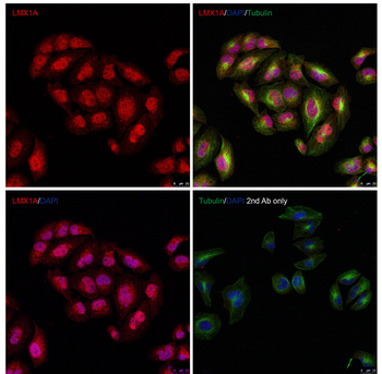
Immunofluorescence Validation of LMX1A in HeLa Cells. Immunofluorescent analysis of methanol-fixed HeLa cells labeling LMX1A with orb1239578 at 10 µg/mL, followed by goat anti-rabbit IgG secondary antibody at 1/1000 dilution (red) and DAPI staining (blue). Alpha tubulin was stained with anti-alpha tubulin antibody following by goat anti-mouse IgG secondary antibody (green). Images were captured with confocal microscopy.
Western Blot Validation in Human Skeletal Muscle. Loading: 10 µg of lysates per lane. Antibodies: LMX1A, orb1239578, 1 µg/mL, 1 h incubation at RT in 5% NFDM/TBST. Secondary: Goat anti-rabbit IgG HRP conjugate at 1:10000 dilution.
Western Blot Validation in Mouse Skeletal Muscle. Loading: 10 µg of lysates per lane. Antibodies: LMX1A, orb1239578, 1 µg/mL, 1 h incubation at RT in 5% NFDM/TBST. Secondary: Goat anti-rabbit IgG HRP conjugate at 1:10000 dilution.
Western Blot Validation in Rat Skeletal Muscle. Loading: 10 µg of lysates per lane. Antibodies: LMX1A, orb1239578, 1 µg/mL, 1 h incubation at RT in 5% NFDM/TBST. Secondary: Goat anti-rabbit IgG HRP conjugate at 1:10000 dilution.
Immunohistochemistry Validation of LMX1A in Human Brain Meningioma. Immunohistochemical analysis of paraffin-embedded human brain meningioma using anti-LMX1A antibody (orb1239578) at 2 µg/mL. Tissue was fixed with formaldehyde and blocked with 10% serum for 1 h at RT, antigen retrieval was by heat mediation with a citrate buffer (pH6). Samples were incubated with primary antibody overnight at 4C. A goat anti-rabbit IgG H&L (HRP) at 1/250 was used as secondary. Counter stained with Hematoxylin.
Immunohistochemistry Validation of LMX1A in Mouse Brain Tissue. Immunohistochemical analysis of paraffin-embedded mouse brain tissue using anti- anti-LMX1A antibody (orb1239578) at 2 µg/mL. Tissue was fixed with formaldehyde and blocked with 10% serum for 1 h at RT, antigen retrieval was by heat mediation with a citrate buffer (pH6). Samples were incubated with primary antibody overnight at 4C. A goat anti-rabbit IgG H&L (HRP) at 1/250 was used as secondary. Counter stained with Hematoxylin.
Immunohistochemistry Validation of LMX1A in Rat Skeletal Muscle Tissue. Immunohistochemical analysis of paraffin-embedded rat spleen tissue using anti-LMX1A antibody (orb1239578) at 5 µg/mL. Tissue was fixed with formaldehyde and blocked with 10% serum for 1 h at RT, antigen retrieval was by heat mediation with a citrate buffer (pH6). Samples were incubated with primary antibody overnight at 4C. A goat anti-rabbit IgG H&L (HRP) at 1/250 was used as secondary. Counter stained with Hematoxylin.
* VAT and and shipping costs not included. Errors and price changes excepted