Anti-SARS-CoV-2 (COVID-19) Spike S1 antibody (orb1239995) was raised against a peptide corresponding to 16 amino acids near the amino terminus of SARS-CoV-2 (COVID-19) Spike S1 glycoprotein. The immunogen is located within the first 50 amino acids of SARS-CoV-2 (COVID-19) Spike S1 protein.
SARS-CoV-2 (COVID-19) Spike S1 antibody is supplied in PBS containing 0.02% sodium azide.
Form:
Liquid
Target:
S
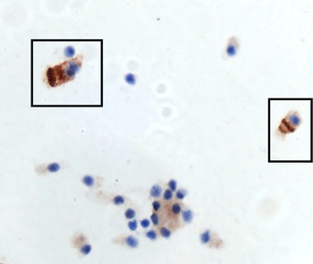
Immunohistochemistry Validation of Spike in the Nasal Swab Sample of Omicron Variant COVID-19 Patient. Immunohistochemical analysis of 4% paraformaldehyde-fixed patient nasal swab sample using spike S1 antibody (orb1239995) at 0.5 µg/ml. Tissue was fixed with formaldehyde and blocked with 10% serum for 1 h at RT, antigen retrieval was by heat mediation with a citrate buffer (pH6). Samples were incubated with primary antibody overnight at 4C. A goat anti-rabbit IgG H&L (HRP) at 1/250 was used as secondary. Counter stained with Hematoxylin. Strong spike protein signal was observed in the nasal swab sample of the Omicron variant COVID-19 patient.
Immunohistochemistry Validation of Spike in Delta Variant COVID-19 Patient Lung Tissue. Immunohistochemical analysis of paraffin-embedded patient lung tissue using anti-Spike S1 antibody (orb1239995) at 0.5 µg/ml. Tissue was fixed with formaldehyde and blocked with 10% serum for 1 h at RT, antigen retrieval was by heat mediation with a citrate buffer (pH6). Samples were incubated with primary antibody overnight at 4C. A goat anti-rabbit IgG H&L (HRP) at 1/250 was used as secondary. Counter stained with Hematoxylin. Strong spike protein signal was observed in the Delta variant COVID-19 patient lung, but not in non-COVID-19 patient lung.
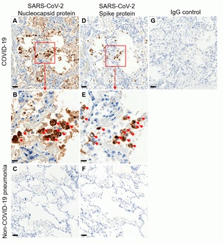
Immunohistochemistry Validation of SARS-CoV-2 (COVID-19) Spike S1 in Human Lung Tissue from the COVID-19 Patient (Sun et al., 2020). Detection of SARS-CoV-2 nucleocapsid protein by anti-SARS-COV-2 nucleocapsid antibodies (orb1239972, 0.02 µg/mL, A, B) or SARS-CoV-2 Spike S1 antibodies (orb1239995, 1 µg/mL D, E) in adjacent sections of autopsy lung tissue from COVID-19 deceased patient. Negative control staining on autopsy lung tissue from patient who died from non-COVID-19 pneumonia is shown for Nucleocapsid protein (C) or Spike protein (F). Negative control using normal rabbit immunoglobulin on COVID-19 autopsy tissue is presented (G). DAB chromogen and hematoxylin counterstain are used. Scale bars: 50µM in A, C, D, F, G, 20µM in B and E.
Immunohistochemistry Validation of SARS-CoV-2 (COVID-19) Spike S1 in COVID-19 Patient Lung. Immunohistochemical analysis of paraffin-embedded COVID-19 patient lung tissue using anti-SARS-CoV-2 (COVID-19) Spike S1 antibody (orb1239995, 0.5 µg/mL). Tissue was fixed with formaldehyde and blocked with 10% serum for 1 h at RT, antigen retrieval was by heat mediation with a citrate buffer (pH6). Samples were incubated with primary antibody overnight at 4 C. A goat anti-rabbit IgG H&L (HRP) at 1/250 was used as secondary. Counter stained with Hematoxylin. Strong signal of SARS-COV-2 spike protein was observed in macrophage of COVID-19 patient lung, but not in non-COVID-19 patient lung.
Immunohistochemistry Validation of SARS-CoV-2 (COVID-19) Spike S1 in Human Lung Tissue from the COVID-19 Patient. Immunohistochemical analysis of paraffin-embedded COVID-19 patient lung tissue using anti-SARS-CoV-2 (COVID-19) Spike S1 antibody (orb1239995, 1 µg/mL). Tissue was fixed with formaldehyde and blocked with 10% serum for 1 h at RT, antigen retrieval was by heat mediation with a citrate buffer (pH6). Samples were incubated with primary antibody overnight at 4C. A goat anti-rabbit IgG H&L (HRP) at 1/250 was used as secondary. Counter stained with Hematoxylin. (Courtesy of Hallgeir Rui, MCW) (Picture shown in 40X magnification).
Overexpression Validation in Spike Transfected 293 Cells. Loading: 10 µg per lane of 293 cell lysate. Antibodies: SARS-CoV-2 (COVID-19) Spike S1, orb1239995 (1 µg/mL), 1h incubation at RT in 5% NFDM/TBST. Secondary: Goat anti-rabbit IgG HRP conjugate at 1:10000 dilution. Lane 1: WT 293 cells and Lane 2: SARS-CoV-2 Spike overexpressed 293 cells.
Immunofluorescence Validation of SARS-CoV-2 (COVID-19) Spike S1 in 293 Transfected Cells. Immunofluorescent analysis of 4% paraformaldehyde-fixed Spike transfected 293 cells labeling SARS-CoV-2 (COVID-19) Spike S1 with orb1239995 at 5 ug/mL, followed by goat anti-rabbit
* VAT and and shipping costs not included. Errors and price changes excepted