Anti-TIM-1 antibody (orb1240149) was raised against a peptide corresponding to 16 amino acids near the amino terminus of human TIM-1. The immunogen is located within amino acids 50 - 100 of TIM-1.
TIM-1 Antibody is supplied in PBS containing 0.02% sodium azide.
Form:
Liquid
Target:
HAVCR1
Application Notes:
Application Notes: WB: 1 - 8 µg/mL (overnight incubation at 4° C), IHC-P: 10 µg/mL, IF: 20 µg/mL. Antibody validated: Western Blot in human and mouse samples, Immunohistochemistry and Immunofluorescence in human samples. All other applications and species not yet tested
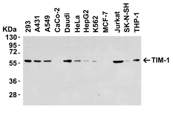
Western Blot Validation in Human Cell Lines. Loading: 15 µg of lysates per lane. Antibodies: TIM-1 orb1240149 (8 µg/mL), overnight incubation at 4C in 5% NFDM/TBST. Secondary: Goat anti-rabbit IgG HRP conjugate at 1:10000 dilution.
Independent Antibody
Independent Antibody Validation (IAV) via Protein Expression Profile in Cell Lines. Loading: 15 µg of lysates per lane. Antibodies: TIM-1 orb1240149 (8 µg/mL), TIM-1 orb1238784 (1 µg/mL), beta-actin (1 µg/mL), and GAPDH (0.02 µg/mL), overnight incubation at 4C (orb1240149) or 1h incubation at RT in 5% NFDM/TBST. Secondary: Goat anti-rabbit IgG HRP conjugate at 1:10000 dilution.
Validation with TIM-1 siRNA Knockdown in Hela Cells. HeLa cells were transfected with control siRNAs (lane 1) or TIM-1 siRNAs (lane 2). Loading: 15 µg of HeLa whole cell lysates per lane. Antibodies: orb1240149 (8 µg/mL), 1 h incubation at RT in 5% NFDM/TBST. Secondary: Goat anti-rabbit IgG HRP conjugate at 1:10000 dilution.
Western Blot Validation in Mouse Tissues. Loading: 15 µg of lysates. Antibodies: TIM-1 orb1240149, 2 µg/mL, 1h incubation at RT in 5% NFDM/TBST. Secondary: Goat anti-rabbit IgG HRP conjugate at 1:10000 dilution.
Immunofluorescence Validation of TIM-1 in Human Testis. Immunofluorescent analysis of 4% paraformaldehyde-fixed human testis tissue labeling TIM-1 with orb1240149 at 10 µg/mL, followed by goat anti-rabbit IgG secondary antibody at 1/500 dilution (red) and DAPI staining (blue).
Immunofluorescence Validation of TIM-1 in Mouse Kidney. Immunofluorescent analysis of 4% paraformaldehyde-fixed mouse kidney tissue labeling TIM-1 with orb1240149 at 10 µg/mL, followed by goat anti-rabbit IgG secondary antibody at 1/500 dilution (red) and DAPI staining (blue).
* VAT and and shipping costs not included. Errors and price changes excepted