
ASPSCR1 Antibody, Unconjugated, Rabbit, Polyclonal
Catalog Number:
BYT-ORB1256361
- Images (9)


| Article Name: | ASPSCR1 Antibody, Unconjugated, Rabbit, Polyclonal |
| Biozol Catalog Number: | BYT-ORB1256361 |
| Supplier Catalog Number: | orb1256361 |
| Alternative Catalog Number: | BYT-ORB1256361-100 |
| Manufacturer: | Biorbyt |
| Host: | Rabbit |
| Category: | Antikörper |
| Application: | IF, IHC, IP, WB |
| Species Reactivity: | Human, Mouse, Rat |
| Immunogen: | Recombinant fusion protein containing a sequence corresponding to amino acids 284-553 of human ASPSCR1 (NP_076988.1). |
| Conjugation: | Unconjugated |
| Alternative Names: | Tether containing UBX domain for GLUT4, Alveolar soft part sarcoma chromosomal region candidate gene 1 protein, Alveolar soft part sarcoma locus, Renal papillary cell carcinoma protein 17, UBX domain-containing protein 9, ASPSCR1, ASPL, RCC17, TUG, UBXD9, UBXN9 |
| ASPSCR1 Antibody |
| Clonality: | Polyclonal |
| Concentration: | batch dependent |
| Molecular Weight: | Observed: 80kDa |
| UniProt: | Q9BZE9 |
| Buffer: | PBS with 0.02% sodium azide, 50% glycerol, pH 7.3. |
| Form: | Liquid |
| Target: | ASPSCR1 |
| Application Notes: | Application Notes: WB: 1:500 - 1:2000IHC: 1:50 - 1:200IF: 1:50 - 1:200IP: 1:50 - 1:100 |
|
|
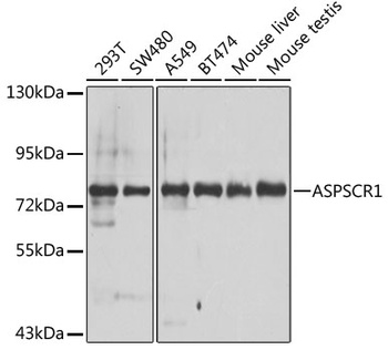
Western blot analysis of extracts of various cell lines, using ASPSCR1 antibody (orb1256361) at 1:1000 dilution. Secondary antibody: HRP Goat Anti-Rabbit IgG (H+L) at 1:10000 dilution. Lysates/proteins: 25 ug per lane. Blocking buffer: 3% nonfat dry milk in TBST. Detection: ECL Basic Kit. Exposure time: 30s. |
|
|
Immunohistochemistry of paraffin-embedded rat kidney using ASPSCR1 antibody (orb1256361) at dilution of 1:100 (40x lens). |
|
|
Immunohistochemistry of paraffin-embedded human kidney cancer using ASPSCR1 antibody (orb1256361) at dilution of 1:100 (40x lens). |
|
|
Immunohistochemistry of paraffin-embedded mouse lung using ASPSCR1 antibody (orb1256361) at dilution of 1:100 (40x lens). |
|
|
Immunohistochemistry of paraffin-embedded mouse kidney using ASPSCR1 antibody (orb1256361) at dilution of 1:100 (40x lens). |
|
|
Immunofluorescence analysis of HeLa cells using ASPSCR1 antibody (orb1256361) at dilution of 1:100 (40x lens). Blue: DAPI for nuclear staining. |
|
|
Immunofluorescence analysis of NIH/3T3 cells using ASPSCR1 antibody (orb1256361) at dilution of 1:100 (40x lens). Blue: DAPI for nuclear staining. |
|
|
Immunofluorescence analysis of PC-12 cells using ASPSCR1 antibody (orb1256361) at dilution of 1:100 (40x lens). Blue: DAPI for nuclear staining. |
|
|
Immunoprecipitation analysis of 200 ug extracts of A-549 cells, using 3 ug ASPSCR1 antibody (orb1256361). Western blot was performed from the immunoprecipitate using ASPSCR1 antibody (orb1256361) at a dilution of 1:1000. |
Product Guarantee and Expert Support
