
H3K18ac Antibody, Unconjugated, Rabbit, Polyclonal
Catalog Number:
BYT-ORB1256501
- Images (11)


| Article Name: | H3K18ac Antibody, Unconjugated, Rabbit, Polyclonal |
| Biozol Catalog Number: | BYT-ORB1256501 |
| Supplier Catalog Number: | orb1256501 |
| Alternative Catalog Number: | BYT-ORB1256501-100 |
| Manufacturer: | Biorbyt |
| Host: | Rabbit |
| Category: | Antikörper |
| Application: | ChIP, IF, IHC, IP, WB |
| Species Reactivity: | Mouse, Rat |
| Immunogen: | A synthetic acetylated peptide corresponding to residues surrounding K18 of human H3 |
| Conjugation: | Unconjugated |
| Alternative Names: | H3/b, H3FB, ERBB, ERBB1, HER1, H3/a, H3/c, H3/d, H3/f, H3/h |
| H3K18ac Antibody |
| Clonality: | Polyclonal |
| Concentration: | batch dependent |
| Molecular Weight: | Observed: 16kDa |
| UniProt: | P68431 |
| Buffer: | PBS with 0.02% sodium azide, 50% glycerol, pH 7.3. |
| Form: | Liquid |
| Target: | H3K18ac |
| Application Notes: | Application Notes: WB: 1:1000 - 1:3000IHC: 1:200 - 1:500IF: 1:500 - 1:1000IP: 1:200 - 1:500CHIP: 1:50 - 1:100CHIP seq: 1:50 - 1:100 |
|
|
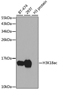
Western blot analysis of extracts of various cell lines, using Acetyl-Histone H3-K18 antibody (orb1256501) at 1:1000 dilution. Secondary antibody: HRP Goat Anti-Rabbit IgG (H+L) at 1:10000 dilution. Lysates/proteins: 25 ug per lane. Blocking buffer: 3% nonfat dry milk in TBST. Detection: ECL Basic Kit. Exposure time: 90s. |
|
|
Immunohistochemistry of paraffin-embedded human stomach using LC3B antibody (orb1274430) at dilution of 1:100 (40x lens). |
|
|
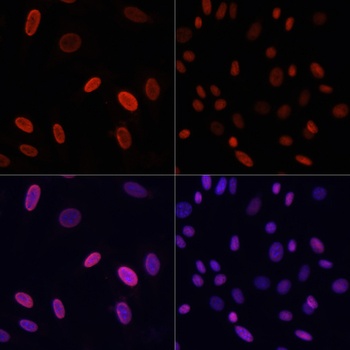
Immunofluorescence analysis of NIH/3T3 cells using Acetyl-Histone H3-K18 antibody (orb1256501) at dilution of 1:100. NIH/3T3 cells were treated by TSA (1 uM) at 37 °C for 18 hours. Blue: DAPI for nuclear staining. |
|
|
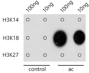
Dot-blot analysis of all sorts of methylation peptides using Acetyl-Histone H3-K18 antibody (orb1256501) at 1:1000 dilution. |
|
|
Immunohistochemistry of paraffin-embedded rat ovary using H3K18ac antibody (orb1256501) at dilution of 1:100 (40x lens). |
|
|
Immunohistochemistry of paraffin-embedded rat brain using H3K18ac antibody (orb1256501) at dilution of 1:100 (40x lens). |
|
|
Immunohistochemistry of paraffin-embedded human stomach using H3K18ac antibody (orb1256501) at dilution of 1:100 (40x lens). |
|
|
Immunohistochemistry of paraffin-embedded human gastric cancer using H3K18ac antibody (orb1256501) at dilution of 1:100 (40x lens). |
|
|
Immunohistochemistry of paraffin-embedded mouse liver using H3K18ac antibody (orb1256501) at dilution of 1:100 (40x lens). |
|
|
Immunohistochemistry of paraffin-embedded mouse brain using H3K18ac antibody (orb1256501) at dilution of 1:100 (40x lens). |
|
|
Immunofluorescence analysis of C6 cells using Acetyl-Histone H3-K18 antibody (orb1256501) at dilution of 1:100. C6 cells were treated by TSA (1 uM) at 37 °C for 18 hours. Blue: DAPI for nuclear staining. |
Product Guarantee and Expert Support
