
ATP5A1 Antibody, Unconjugated, Rabbit, Polyclonal
Catalog Number:
BYT-ORB1257272
- Images (9)


| Article Name: | ATP5A1 Antibody, Unconjugated, Rabbit, Polyclonal |
| Biozol Catalog Number: | BYT-ORB1257272 |
| Supplier Catalog Number: | orb1257272 |
| Alternative Catalog Number: | BYT-ORB1257272-100 |
| Manufacturer: | Biorbyt |
| Host: | Rabbit |
| Category: | Antikörper |
| Application: | IF, IHC, IP, WB |
| Species Reactivity: | Human, Mouse, Rat |
| Immunogen: | Recombinant fusion protein containing a sequence corresponding to amino acids 1-280 of human ATP5A1 (NP_004037.1). |
| Conjugation: | Unconjugated |
| Alternative Names: | ATP5A1, ATP synthase, H+ transporting, mitochondrial F1 complex, alpha subunit 1, cardiac muscle, ATP5A, ATP5AL2, ATPM, MOM2, OMR, ORM, hATP1, ATP synthase alpha chain, mitochondrial, ATP synthase subunit alpha, mitochondrial, ATP synthase, H+ transportin |
| ATP5A1 Antibody |
| Clonality: | Polyclonal |
| Concentration: | batch dependent |
| Molecular Weight: | Observed: 53kDa |
| UniProt: | P25705 |
| Buffer: | PBS with 0.02% sodium azide, 50% glycerol, pH 7.3. |
| Form: | Liquid |
| Target: | ATP5A1 |
| Application Notes: | Application Notes: WB: 1:500 - 1:2000IHC: 1:50 - 1:200IF: 1:50 - 1:100IP: 1:50 - 1:200 |
|
|
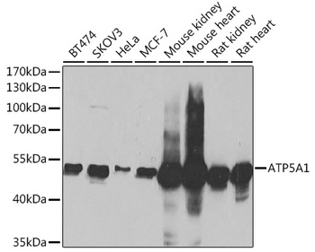
Western blot analysis of extracts of various cell lines, using ATP5A1 Antibody (orb1257272) at 1:1000 dilution. Secondary antibody: HRP Goat Anti-Rabbit IgG (H+L) at 1:10000 dilution. Lysates/proteins: 25 ug per lane. Blocking buffer: 3% nonfat dry milk in TBST. Detection: ECL Basic Kit. Exposure time: 1s. |
|
|
Immunohistochemistry of paraffin-embedded rat ovary using ATP5A1 antibody (orb1257272) at dilution of 1:100 (40x lens). |
|
|
Immunohistochemistry of paraffin-embedded human liver cancer using ATP5A1 antibody (orb1257272) at dilution of 1:100 (40x lens). |
|
|
Immunohistochemistry of paraffin-embedded human stomach using ATP5A1 antibody (orb1257272) at dilution of 1:100 (40x lens). |
|
|
Immunohistochemistry of paraffin-embedded mouse liver using ATP5A1 antibody (orb1257272) at dilution of 1:100 (40x lens). |
|
|
Immunofluorescence analysis of HeLa cells using ATP5A1 antibody (orb1257272) at dilution of 1:100 (60x lens). Blue: DAPI for nuclear staining. |
|
|
Confocal immunofluorescence analysis of Hela cells using ATP5A1 antibody (orb1257272) at dilution of 1:200. Blue: DAPI for nuclear staining. |
|
|
Confocal immunofluorescence analysis of U-2OS cells using ATP5A1 antibody (orb1257272) at dilution of 1:200. Blue: DAPI for nuclear staining. |
|
|
Immunoprecipitation analysis of 200 ug extracts of MCF-7 cells using 1 ug ATP5A1 antibody (orb1257272). Western blot was performed from the immunoprecipitate using ATP5A1 antibody (orb1257272) at a dilution of 1:1000. |
Product Guarantee and Expert Support
