
H3K9me1 Antibody, Unconjugated, Rabbit, Polyclonal
Catalog Number:
BYT-ORB1258190
- Images (9)


| Article Name: | H3K9me1 Antibody, Unconjugated, Rabbit, Polyclonal |
| Biozol Catalog Number: | BYT-ORB1258190 |
| Supplier Catalog Number: | orb1258190 |
| Alternative Catalog Number: | BYT-ORB1258190-100 |
| Manufacturer: | Biorbyt |
| Host: | Rabbit |
| Category: | Antikörper |
| Application: | ChIP, IF, IHC, IP, WB |
| Species Reactivity: | Human, Mouse, Other, Rat |
| Immunogen: | A synthetic methylated peptide corresponding to residues surrounding K9 of human histone H3 |
| Conjugation: | Unconjugated |
| Alternative Names: | H3F3A, H3t, H3.4, H3/g, H3FT |
| H3K9me1 Antibody |
| Clonality: | Polyclonal |
| Concentration: | batch dependent |
| Molecular Weight: | Observed: 18kDa |
| UniProt: | Q16695 |
| Buffer: | PBS with 0.02% sodium azide, 50% glycerol, pH 7.3. |
| Form: | Liquid |
| Target: | H3K9me1 |
| Application Notes: | Application Notes: WB: 1:500 - 1:2000IHC: 1:50 - 1:200IF: 1:50 - 1:200IP: 1:50 - 1:200ChIP: 1:20 - 1:100CHIP seq: 1:20 - 1:100 |
|
|
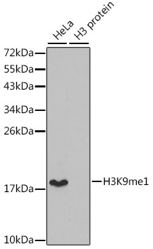
Western blot analysis of extracts of various cell lines, using MonoMethyl-Histone H3-K9 antibody (orb1258190). Secondary antibody: HRP Goat Anti-Rabbit IgG (H+L) at 1:10000 dilution. Lysates/proteins: 25 ug per lane. Blocking buffer: 3% nonfat dry milk in TBST. |
|
|
Dot-blot analysis of all sorts of methylation peptides using MonoMethyl-Histone H3-K9 antibody (orb1258190). |
|
|
Immunohistochemistry of paraffin-embedded rat testis using MonoMethyl-Histone H3-K9 antibody (orb1258190) at dilution of 1:200 (40x lens). |
|
|
Immunohistochemistry of paraffin-embedded human embryo brain using MonoMethyl-Histone H3-K9 antibody (orb1258190) at dilution of 1:200 (40x lens). |
|
|
Immunofluorescence analysis of 293T cells using MonoMethyl-Histone H3-K9 antibody (orb1258190). Blue: DAPI for nuclear staining. |
|
|
Immunofluorescence analysis of C6 cells using MonoMethyl-Histone H3-K9 antibody (orb1258190) at dilution of 1:100. Blue: DAPI for nuclear staining. |
|
|
Immunofluorescence analysis of HeLa cells using MonoMethyl-Histone H3-K9 antibody (orb1258190) at dilution of 1:100. Blue: DAPI for nuclear staining. |
|
|
Immunofluorescence analysis of NIH/3T3 cells using MonoMethyl-Histone H3-K9 antibody (orb1258190) at dilution of 1:100. Blue: DAPI for nuclear staining. |
|
|
Chromatin immunoprecipitation analysis of extracts of 293 cell line, using MonoMethyl-Histone H3-K9 antibody (orb1258190) and rabbit IgG. The amount of immunoprecipitated DNA was checked by quantitative PCR. Histogram was constructed by the ratios of the immunoprecipitated DNA to the input. |
Product Guarantee and Expert Support
