
CTGF Antibody, Unconjugated, Rabbit, Polyclonal
Catalog Number:
BYT-ORB1261312
- Images (9)


| Article Name: | CTGF Antibody, Unconjugated, Rabbit, Polyclonal |
| Biozol Catalog Number: | BYT-ORB1261312 |
| Supplier Catalog Number: | orb1261312 |
| Alternative Catalog Number: | BYT-ORB1261312-100 |
| Manufacturer: | Biorbyt |
| Host: | Rabbit |
| Category: | Antikörper |
| Application: | IF, IHC, WB |
| Species Reactivity: | Human, Mouse, Rat |
| Immunogen: | Recombinant fusion protein containing a sequence corresponding to amino acids 27-349 of human CTGF (NP_001892.1). |
| Conjugation: | Unconjugated |
| Alternative Names: | CCN2, NOV2, HCS24, IGFBP8, CCN2, Connective tissue growth factor, CCN family member 2, IBP-8 |
| CTGF Antibody |
| Clonality: | Polyclonal |
| Concentration: | batch dependent |
| Molecular Weight: | Observed: 38kDa |
| UniProt: | P29279 |
| Buffer: | PBS with 0.02% sodium azide, 50% glycerol, pH 7.3. |
| Form: | Liquid |
| Target: | CTGF |
| Application Notes: | Application Notes: WB: 1:500 - 1:2000IHC: 1:50 - 1:200IF: 1:20 - 1:50 |
|
|
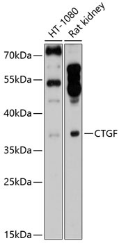
Western blot analysis of extracts of various cell lines, using CTGF antibody (orb1261312) at 1:1000 dilution. Secondary antibody: HRP Goat Anti-Rabbit IgG (H+L) at 1:10000 dilution. Lysates/proteins: 25 ug per lane. Blocking buffer: 3% nonfat dry milk in TBST. Detection: ECL Enhanced Kit. Exposure time: 30s. |
|
|
Immunohistochemistry of paraffin-embedded rat heart using CTGF antibody (orb1261312) at dilution of 1:100 (40x lens). |
|
|
Immunohistochemistry of paraffin-embedded human breast using CTGF antibody (orb1261312) at dilution of 1:100 (40x lens). |
|
|
Immunohistochemistry of paraffin-embedded mouse kidney using CTGF antibody (orb1261312) at dilution of 1:100 (40x lens). |
|
|
Immunofluorescence analysis of C6 cells using CTGF antibody (orb1261312) at dilution of 1:100. Blue: DAPI for nuclear staining. |
|
|
Immunofluorescence analysis of NIH/3T3 cells using CTGF antibody (orb1261312) at dilution of 1:100. Blue: DAPI for nuclear staining. |
|
|
Immunofluorescence analysis of U2OS cells using CTGF antibody (orb1261312) at dilution of 1:100. Blue: DAPI for nuclear staining. |
|
|
Immunofluorescence analysis of U2OS cells using CTGF antibody (orb1261312) at dilution of 1:100. Blue: DAPI for nuclear staining. |
|
|
Immunofluorescence analysis of HeLa cells using CTGF antibody (orb1261312) at dilution of 1:100 (40x lens). Blue: DAPI for nuclear staining. |
Product Guarantee and Expert Support
