Synthetic peptide / Extracellular domain corresponding to Human ICAM-1 / CD54
Conjugation:
Unconjugated
Rabbit polyclonal antibody to ICAM-1
Clonality:
Polyclonal
Target:
ICAM-1 / CD54 (Extracellular)
Application Dilute:
Western Blot 1:500 Immunofluorescence 1:200-1:500 Immunohistochemistry (Paraffin) 1:100-1:200
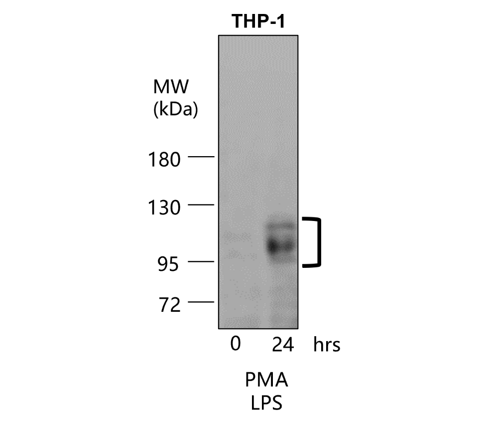
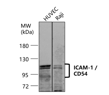
ICAM-1 / CD54 antibody - at 1/500 dilution, Lysates/proteins at 30 µg per lane. This blot was produced using a 7.5% SDS-PAGE. Nitrocellulose membrane was then blocked with 3% BSA for an hour before being incubated with orb1294365 overnight at 4C.
ICAM-1 / CD54 antibody - at 1/500 dilution, Lysates/proteins at 30 µg per lane. This blot was produced using a 7.5% SDS-PAGE. Nitrocellulose membrane was then blocked with 3% BSA for an hour before being incubated with orb1294365 overnight at 4C.
ICAM-1 / CD54 antibody - at 1/500 dilution, Lysates/proteins at 30 µg per lane. This blot was produced using a 7.5% SDS-PAGE. Nitrocellulose membrane was then blocked with 3% BSA for an hour before being incubated with orb1294365 overnight at 4C.
Immunofluorescence: cells were fixed with 4% paraformaldehyde for 10 min at RT, permeabilized with 0.1% NP-40 for 10 min at RT then blocked with 5% BSA for 30 min at room temperature. Cells were stained with orb1294365 anti-ICAM-1 antibody (red) at 1:100 and 4C. DAPI (blue) was used as the nuclear counter stain.
Immunofluorescence: cells were fixed with 4% paraformaldehyde for 10 min at RT, permeabilized with 0.1% NP-40 for 10 min at RT then blocked with 5% BSA for 30 min at room temperature. Cells were stained with orb1294365 anti-ICAM-1 antibody (red) at 1:100 and 4C. DAPI (blue) was used as the nuclear counter stain.
Immunohistochemical analysis of paraffin embedded Human cancer tissue labeling ICAM-1 at 1/100.
* VAT and and shipping costs not included. Errors and price changes excepted