
GFAP Rabbit Polyclonal Antibody, Unconjugated
Catalog Number:
BYT-ORB1294403
- Images (8)


| Article Name: | GFAP Rabbit Polyclonal Antibody, Unconjugated |
| Biozol Catalog Number: | BYT-ORB1294403 |
| Supplier Catalog Number: | orb1294403 |
| Alternative Catalog Number: | BYT-ORB1294403-25,BYT-ORB1294403-100 |
| Manufacturer: | Biorbyt |
| Host: | Rabbit |
| Category: | Antikörper |
| Application: | IF, IHC-Fr, IHC-P, WB |
| Species Reactivity: | Human, Mouse, Rat |
| Immunogen: | Synthetic peptide / encompassing a sequence within the C-terminus region. |
| Conjugation: | Unconjugated |
| Rabbit polyclonal antibody to GFAP |
| Clonality: | Polyclonal |
| Buffer: | 100mM Tris Glycine, 1% rAlbumin, 20% Glycerol (pH7). 0.025% ProClin 300 was added as a preservative |
| Target: | GFAP |
| Application Dilute: | Western Blot 1:1000-1:2000, Immunofluorescence 1:200 - 1:500, Immunohistochemistry (Frozen) 1:100 - 1:300, Immunohistochemistry (Paraffin) 1:100 - 1:300 |
|
|
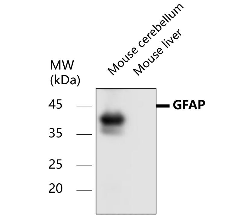
All lanes: Anti-GFAP antibody at 1/1000 dilution, Lysates/proteins at 60 µg per lane. This blot was produced using a 12% SDS-PAGE. Nitrocellulose was then blocked for an hour before being incubated with orb1294403 overnight at 4C. |
|
|
All lanes: Anti-GFAP antibody at 1/1000 dilution, Lysates/proteins at 60 µg per lane. This blot was produced using a 12% SDS-PAGE. Nitrocellulose was then blocked for an hour before being incubated with orb1294403 overnight at 4C. |
|
|
Immunofluorescence: cells were fixed with 4% paraformaldehyde for 10 min at RT, permeabilized with 0.1% NP-40 for 10 min at RT then blocked with 5% BSA for 30 min at room temperature. Cells were stained with orb1294403 anti-GFAP antibody (red) at 1:200 and 4C. DAPI (blue) was used as the nuclear counter stain. |
|
|
Immunofluorescent analysis. Sample: primary cortical neurons, Red: GFAP (orb1294403): 1-200, Blue: DAPI was used as the nuclear counter stain. Fixed: 4% paraformaldehyde at RT for 20 min. |
|
|
Immunohistochemical analysis of paraffin embedded Human brain tissue labeling GFAP antibody with orb1294403 at 1/100. |
|
|
Immunohistochemical analysis of paraffin embedded Mouse brain tissue labeling GFAP antibody with orb1294403 at 1/100. |
|
|
Immunohistochemical of frozen sections. Sample: mouse cerebellum. Green: GFAP (orb1294403): 1-200, Anti-rabbit 488: 1-500. |
|
|
Immunohistochemical of frozen sections. Sample: mouse cerebellum. Green: GFAP (orb1294403): 1-200, Anti-rabbit 488: 1-500. |
Product Guarantee and Expert Support
