HSPD1, HSP60, 60 kDa heat shock protein, mitochondrial, Chaperonin 60, CPN60, HuCHA60, Heat shock protein family D member 1, GroEL homolog, mitochondrial, GROEL, HLD4, HSP 60, HSP65, SPG 13
Mouse monoclonal to Hsp60 (Biotin). In both prokaryotic and eukaryotic cells, the misfolding In both prokaryotic and eukaryotic cells, the misfolding...
136.36mM Ethanolamine, 133.23 mM Chlorides, 9.55mM Phosphates, 9.55mM Sodium Bicarbonate
Target:
HSP60
Application Dilute:
WB (1:20000), IHC (1:100), ICC/IF (1:100), IP (1:200)
Application Notes:
Application Notes: 0.05 µg/ml of SMC-110 was sufficient for detection of HSP60 in 20 µg of heat shocked HeLa cell lysate by colorimetric immunoblot analysis using goat anti-mouse IgG as the secondary antibody
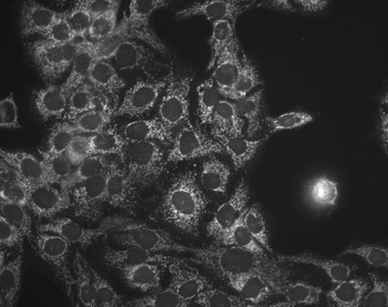
Immunocytochemistry/Immunofluorescence analysis using Mouse Anti-Hsp60 Monoclonal Antibody, Clone LK-1. Tissue: HaCaT cells. Species: Human. Fixation: Cold 100% methanol at -20C for 10 minutes. Primary Antibody: Mouse Anti-Hsp60 Monoclonal Antibody at 1:100 for 1 hour at RT. Secondary Antibody: FITC Goat Anti-Mouse (green) at 1:50 for 1 hour at RT. Localization: Cytoplasmic Staining.
Immunohistochemistry analysis using Mouse Anti-Hsp60 Monoclonal Antibody, Clone LK-1. Tissue: colon carcinoma. Species: Human. Fixation: Formalin. Primary Antibody: Mouse Anti-Hsp60 Monoclonal Antibody at 1:100000 for 12 hours at 4C. Secondary Antibody: Biotin Goat Anti-Mouse at 1:2000 for 1 hour at RT. Counterstain: Mayer Hematoxylin (purple/blue) nuclear stain at 200 µl for 2 minutes at RT. Localization: Inflammatory cells. Magnification: 40x.
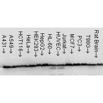
Western Blot analysis of Human Cell line lysates showing detection of Hsp60 protein using Mouse Anti-Hsp60 Monoclonal Antibody, Clone LK-1. Load: 15 µg. Block: 1.5% BSA for 30 minutes at RT. Primary Antibody: Mouse Anti-Hsp60 Monoclonal Antibody at 1:1000 for 2 hours at RT. Secondary Antibody: Sheep Anti-Mouse IgG: HRP for 1 hour at RT.
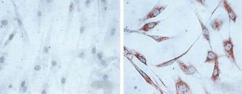
Immunocytochemistry/Immunofluorescence analysis using Mouse Anti-Hsp60 Monoclonal Antibody, Clone LK1, . Tissue: skin Fibroblasts. Species: Human. Fixation: Cold 100% methanol for 30 minutes at -20C. Primary Antibody: Mouse Anti-Hsp60 Monoclonal Antibody at 1:1000 for 1 hour at RT. Secondary Antibody: DAKO LSAB2 streptavidin-peroxidase system. Counterstain: Mayer Hematoxylin (purple/blue) nuclear stain. Left: control, Right: 24 hours after 7th passage of senescence.
Immunohistochemistry analysis using Mouse Anti-Hsp60 Monoclonal Antibody, Clone LK-1. Tissue: backskin. Species: Mouse. Fixation: Bouins Fixative and paraffin-embedded. Primary Antibody: Mouse Anti-Hsp60 Monoclonal Antibody at 1:100 for 1 hour at RT. Secondary Antibody: FITC Goat Anti-Mouse (green) at 1:50 for 1 hour at RT. Localization: Epidermis.
* VAT and and shipping costs not included. Errors and price changes excepted