136.36mM Ethanolamine, 133.23 mM Chlorides, 9.55mM Phosphates, 9.55mM Sodium Bicarbonate
Target:
PSD95
Application Dilute:
WB (1:1000), IHC (1:1000), ICC/IF (1:100)
Application Notes:
Application Notes: 1 µg/ml was sufficient for detection of PSD-95 on 20 µg rat brain tissue extract by ECL immunoblot analysis using Goat Anti-Mouse IgG: HRP as the secondary
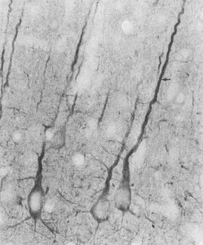
Immunohistochemistry analysis using Mouse Anti-PSD95 Monoclonal Antibody, Clone 7E3. Tissue: Neocortex. Species: Rat. Primary Antibody: Mouse Anti-PSD95 Monoclonal Antibody at 1:1000.
Immunocytochemistry/Immunofluorescence analysis using Mouse Anti-PSD95 Monoclonal Antibody, Clone 7E3. Tissue: HaCaT cells. Species: Human. Fixation: Cold 100% methanol for 10 minutes at -20C. Primary Antibody: Mouse Anti-PSD95 Monoclonal Antibody at 1:100 for 1 hour at RT. Secondary Antibody: FITC Goat Anti-Mouse (green) at 1:50 for 1 hour at RT. Localization: Filamentous-like staining.
Immunohistochemistry analysis using Mouse Anti-PSD95 Monoclonal Antibody, Clone 7E3. Tissue: backskin. Species: Mouse. Fixation: Bouins Fixative and paraffin-embedded. Primary Antibody: Mouse Anti-PSD95 Monoclonal Antibody at 1:100 for 1 hour at RT. Secondary Antibody: FITC Goat Anti-Mouse (green) at 1:50 for 1 hour at RT. Localization: Basal cell staining in the epidermis, some hair follicle staining, dermal staining. Backskin obtained from transgenic mice.
Western Blot analysis of Rat brain membrane lysate showing detection of PSD95 protein using Mouse Anti-PSD95 Monoclonal Antibody, Clone 7E3. Primary Antibody: Mouse Anti-PSD95 Monoclonal Antibody at 1:1000.
* VAT and and shipping costs not included. Errors and price changes excepted