136.36mM Ethanolamine, 133.23 mM Chlorides, 9.55mM Phosphates, 9.55mM Sodium Bicarbonate
Target:
HO-1
Application Dilute:
WB (1:1000), IHC (1:100), ICC/IF (1:100)
Application Notes:
Application Notes: 1 µg/ml was sufficient for detection of HO-1 in 10 µg of mixed human cell line lysate by colorimetric immunoblot analysis using Goat Anti-Mouse IgG:HRP as the secondary
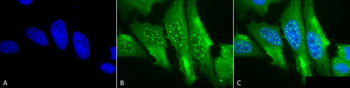
Immunocytochemistry/Immunofluorescence analysis using Mouse Anti-HO-1 Monoclonal Antibody, Clone 1F12-A6. Tissue: Cervical cancer cell line (HeLa). Species: Human. Fixation: 2% Formaldehyde for 20 min at RT. Primary Antibody: Mouse Anti-HO-1 Monoclonal Antibody at 1:100 for 12 hours at 4C. Secondary Antibody: FITC Goat Anti-Mouse (green) at 1:200 for 2 hours at RT. Counterstain: DAPI (blue) nuclear stain at 1:40000 for 2 hours at RT. Localization: Microsome. Endoplasmic reticulum. Localizes to the nucleus upon hypoxia. Magnification: 100x. (A) DAPI (blue) nuclear stain. (B) Anti-HO-1 Antibody. (C) Composite.
Immunohistochemistry analysis using Mouse Anti-HO-1 Monoclonal Antibody, Clone 1F12-A6. Tissue: backskin. Species: Mouse. Fixation: Bouins Fixative and paraffin-embedded. Primary Antibody: Mouse Anti-HO-1 Monoclonal Antibody at 1:100 for 1 hour at RT. Secondary Antibody: FITC Goat Anti-Mouse (green) at 1:50 for 1 hour at RT. Localization: muscle, dermis, hair follicles, epidermis: nuclear everywhere and some cytoplasmic staining.
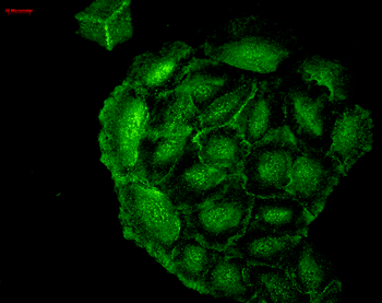
Immunocytochemistry/Immunofluorescence analysis using Mouse Anti-HO-1 Monoclonal Antibody, Clone 1F12-A6. Tissue: Cervical cancer cell line (HeLa). Species: Human. Fixation: 2% Formaldehyde for 20 min at RT. Primary Antibody: Mouse Anti-HO-1 Monoclonal Antibody at 1:100 for 12 hours at 4C. Secondary Antibody: R-PE Goat Anti-Mouse (yellow) at 1:200 for 2 hours at RT. Counterstain: DAPI (blue) nuclear stain at 1:40000 for 2 hours at RT. Localization: Microsome. Endoplasmic reticulum. Localizes to the nucleus upon hypoxia. Magnification: 20x. (A) DAPI (blue) nuclear stain. (B) Anti-HO-1 Antibody. (C) Composite.
Immunocytochemistry/Immunofluorescence analysis using Mouse Anti-HO-1 Monoclonal Antibody, Clone 1F12-A6. Tissue: HaCaT cells. Species: Human. Fixation: Cold 100% methanol for 10 minutes at -20C. Primary Antibody: Mouse Anti-HO-1 Monoclonal Antibody at 1:100 for 1 hour at RT. Secondary Antibody: FITC Goat Anti-Mouse (green) at 1:50 for 1 hour at RT. Localization: Cell-cell border staining in epidermis, punctuate nuclear staining.
Western Blot analysis of Human Cervical cancer cell line (HeLa) lysate showing detection of HO-1 protein using Mouse Anti-HO-1 Monoclonal Antibody, Clone 1F12-A6. Load: 15 µg. Block: 1.5% BSA for 30 minutes at RT. Primary Antibody: Mouse Anti-HO-1 Monoclonal Antibody at 1:1000 for 2 hours at RT. Secondary Antibody: Sheep Anti-Mouse IgG: HRP for 1 hour at RT.
* VAT and and shipping costs not included. Errors and price changes excepted