
ERp57 Antibody (Biotin), IgG1, Clone: [Map.ERp57], Mouse, Monoclonal
Catalog Number:
BYT-ORB147636
- Images (4)


| Article Name: | ERp57 Antibody (Biotin), IgG1, Clone: [Map.ERp57], Mouse, Monoclonal |
| Biozol Catalog Number: | BYT-ORB147636 |
| Supplier Catalog Number: | orb147636 |
| Alternative Catalog Number: | BYT-ORB147636-200 |
| Manufacturer: | Biorbyt |
| Host: | Mouse |
| Category: | Antikörper |
| Application: | ICC, IF, IHC, IP, WB |
| Species Reactivity: | Bovine, Canine, Guinea pig, Hamster, Human, Monkey, Mouse, Porcine, Rabbit, Rat |
| Immunogen: | Human recombinant ERp57 (Grp58) |
| Conjugation: | Biotin |
| Alternative Names: | ERp60, ERp61, Grp57, Grp58, P58, PDIA3, PI PLC, 58 kDa glucose regulated protein, 58 kDa glucose-regulated protein, 58 kDa microsomal protein, Disulfide isomerase ER 60, Disulfide isomerase ER-60, Endoplasmic reticulum resident protein 57, Endoplasmic reticulum resident protein 60, ER p57, ER protein 57, ER protein 60, ERp 57, ERp57, Glucose Regulated Protein 58 Kd, GRP 57, GRP 58, GRP57, HsT17083, p58, PDIA 3, PDIA3_HUMAN, Phospholipase C alpha, Protein disulfide isomerase A3, Protein disulfide isomerase family A member 3, Protein disulfide-isomerase A3 |
| Mouse monoclonal to Erp57 (Biotin). ERp57, also known as Glucose Regulated Protein 58 (Grp58), Hormone-Induced Protein-70 (HIP-70) and microsomal Carnitine Palmitoyltransferase, is a member of the protein disulfide isomerase family, containing two canonical CXHC tetrapeptide active site motifs (1-5). It has quite a few diverse roles. It functions as an accessory oxidoreductase involved in disulfide bond formation. In the ER, ERp57 interacts with membrane bound calnexin and soluble calreticulin (lectin chaperones) via their praline rich P-domain arms. Lectin chaperones bind nascent non-native glycoproteins, and position ERp57 to act upon the immature or misfolded glycoproteins that possess mono-glycosylated side chains. ERp57 deletion impairs posttranslational phases of influenza hema-glutinin folding, and causes accelerated release of MHC-I molecules, resulting in the coupling of sub-optimal peptides and reduced expression and stability on the cell surface. ERp57 also contains two thioredoxin active-site sequences, CGHC and an estrogen-binding domain. ERp57 is induced by both estrogen and leuteinizing-hormone-releasing hormone in the hippocampus.. |
| Application Dilute: | WB (1:2000), IHC (1:100), ICC/IF (1:100) |
| Application Notes: | Application Notes: 0.5 µg/ml of SMC-168 was sufficient for detection of ERp57 in 10 µg of heat shock Heal Lysate by colorimetric immunoblot analysis using Goat anti-mouse IgG:HRP as the secondary antibody |
|
|
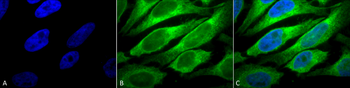
Immunocytochemistry/Immunofluorescence analysis using Mouse Anti-Erp57 (Grp58) Monoclonal Antibody, Clone Map.ERP57. Tissue: Heat Shocked cervical cancer cells (HeLa). Species: Human. Fixation: 2% Formaldehyde for 20 min at RT. Primary Antibody: Mouse Anti-Erp57 (Grp58) Monoclonal Antibody at 1:100 for 12 hours at 4C. Secondary Antibody: FITC Goat Anti-Mouse (green) at 1:200 for 2 hours at RT. Counterstain: DAPI (blue) nuclear stain at 1:40000 for 2 hours at RT. Localization: Endoplasmic reticulum lumen. Melanosome. Magnification: 100x. (A) DAPI (blue) nuclear stain. (B) Anti-Erp57 (Grp58) Antibody. (C) Composite. Heat Shocked at 42C for 1h. |
|
|
Immunohistochemistry analysis using Mouse Anti-Erp57 Monoclonal Antibody, Clone Map.ERP57. Tissue: backskin. Species: Mouse. Fixation: Bouins Fixative and paraffin-embedded. Primary Antibody: Mouse Anti-Erp57 Monoclonal Antibody at 1:100 for 1 hour at RT. Secondary Antibody: FITC Goat Anti-Mouse (green) at 1:50 for 1 hour at RT. Localization: Epidermis and Hair Follicles. |
|
|
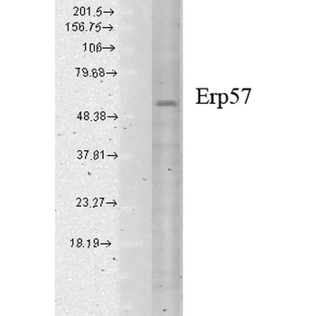
Western Blot analysis of Human cell lysates showing detection of Erp57 protein using Mouse Anti-Erp57 Monoclonal Antibody, Clone Map.ERP57. Load: 15 µg. Block: 1.5% BSA for 30 minutes at RT. Primary Antibody: Mouse Anti-Erp57 Monoclonal Antibody at 1:1000 for 2 hours at RT. Secondary Antibody: Sheep Anti-Mouse IgG: HRP for 1 hour at RT. |
|
|
Immunocytochemistry/Immunofluorescence analysis using Mouse Anti-Erp57 Monoclonal Antibody, Clone Map.ERP57. Tissue: HaCaT cells. Species: Human. Fixation: Cold 100% methanol for 10 minutes at -20C. Primary Antibody: Mouse Anti-Erp57 Monoclonal Antibody at 1:100 for 1 hour at RT. Secondary Antibody: FITC Goat Anti-Mouse (green) at 1:50 for 1 hour at RT. Localization: Cytoplasmic and perinuclear staining. |
Product Guarantee and Expert Support
