AHA1 Antibody (Biotin), Clone: [25F2.D9], Rat, Monoclonal
Biozol Catalog Number:
BYT-ORB147670
Supplier Catalog Number:
orb147670
Alternative Catalog Number:
BYT-ORB147670-100
Manufacturer:
Biorbyt
Host:
Rat
Category:
Antikörper
Application:
ELISA, ICC, IF, IHC, IP, WB
Species Reactivity:
Human, Mouse, Rat
Immunogen:
Recombinant Full Length Mouse Aha1 Protein
Conjugation:
Biotin
Alternative Names:
AHSA1, AHA1, HSPC322, p38, C14orf3, LOC10598, Activator of Hsp90 ATPase activity 1, Activator of 90 kDa heat shock protein ATPase homolog 1
Rat monoclonal to Aha1 (Biotin). Aha1 is a member of the Hsp90 cochaperone family, and is thought to stimulate Hsp90 ATPase activity by competing with p23 and other co-chaperones for Hsp90 binding. It may affect a step in the endoplasmic reticulum to Golgi trafficking. Aha1 also interacts with HSPCA/Hsp90 and with the cytoplasmic tail of the vesicular stomatistis virus glycoproteins (VSV G). Aha1 is expressed in numerous tissues, including the brain, heart, skeletal muscle, and kidney, and at low levels, the liver and placenta. Aha1 might be a potential therapeutic strategy to increase sensitivity to HSP inhibitors..
136.36mM Ethanolamine, 133.23 mM Chlorides, 9.55mM Phosphates, 9.55mM Sodium Bicarbonate
Target:
AHA1
Application Dilute:
WB (1:1000), IHC (1:100), ICC/IF (1:1000), IP (1:1000)
Application Notes:
Application Notes: 1 µg/ml of SMC-172 was sufficient for detection of Aha1 in 10 µg of rat tissue lysate by colorimetric immunoblot analysis using Goat anti-rat IgG:HRP as the secondary antibody
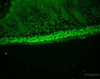
Immunohistochemistry analysis using Rat Anti-Aha1 Monoclonal Antibody, Clone 25F2.D9. Tissue: backskin. Species: Mouse. Fixation: Bouins Fixative and paraffin-embedded. Primary Antibody: Rat Anti-Aha1 Monoclonal Antibody at 1:100 for 1 hour at RT. Secondary Antibody: FITC Goat Anti-Rat (green) at 1:50 for 1 hour at RT. Localization: Uppermost epidermis staining, and muscle.
Immunoprecipitation analysis using Rat Anti-Aha1 Monoclonal Antibody, Clone 25F2.D9. Tissue: HeLa cells. Species: Human. Primary Antibody: Rat Anti-Aha1 Monoclonal Antibody at 1:1000.
Immunocytochemistry/Immunofluorescence analysis using Rat Anti-Aha1 Monoclonal Antibody, Clone 25F2.D9. Tissue: Cervical cancer cell line (HeLa). Species: Human. Primary Antibody: Rat Anti-Aha1 Monoclonal Antibody at 1:1000. Secondary Antibody: FITC Goat Anti-Rat (green).
Western Blot analysis of Human Cell lysates showing detection of Aha1 protein using Rat Anti-Aha1 Monoclonal Antibody, Clone 25F2.D9. Load: 15 µg. Block: 1.5% BSA for 30 minutes at RT. Primary Antibody: Rat Anti-Aha1 Monoclonal Antibody at 1:1000 for 2 hours at RT. Secondary Antibody: Sheep Anti-Mouse IgG: HRP for 1 hour at RT.
* VAT and and shipping costs not included. Errors and price changes excepted