KCNQ4 Antibody (Biotin), IgG1, Clone: [N43/6 Formerly sold as S43-6], Mouse, Monoclonal
Biozol Catalog Number:
BYT-ORB148314
Supplier Catalog Number:
orb148314
Alternative Catalog Number:
BYT-ORB148314-100
Manufacturer:
Biorbyt
Host:
Mouse
Category:
Antikörper
Application:
AM, ICC, IF, IHC, WB
Species Reactivity:
Human, Mouse, Rat
Immunogen:
Fusion protein amino acids 2-77 of human KCNQ4
Conjugation:
Biotin
Alternative Names:
KCNQ4, Kv7.4, KVLQT4, Potassium voltage-gated channel subfamily KQT member 4, Voltage-gated potassium channel subunit Kv7.4, KQT-like 4, Potassium channel subunit alpha KvLQT4, Potassium voltage-gated channel KQT-like subfamily member 4, DFNA2, KCNQ4_HUMAN, Kcnq4, KCNQ 4, Potassium channel KQT like 4, Potassium voltage gated channel KQT like protein 4, Voltage gated potassium channel subunit Kv7.4
Mouse monoclonal to KCNQ4 (Biotin). Ion channels are integral membrane proteins that help establish and control the small voltage gradient across the plasma membrane of living cells by allowing the flow of ions down their electrochemical gradient. They are present in the membranes that surround all biological cells because their main function is to regulate the flow of ions across this membrane. Whereas some ion channels permit the passage of ions based on charge, others conduct based on a ionic species, such as sodium or potassium. Furthermore, in some ion channels, the passage is governed by a gate which is controlled by chemical or electrical signals, temperature, or mechanical forces. There are a few main classifications of gated ion channels. There are voltage- gated ion channels, ligand- gated, other gating systems and finally those that are classified differently, having more exotic characteristics. The first are voltage- gated ion channels which open and close in response to membrane potential. These are then separated into sodium, calcium, potassium, proton, transient receptor, and cyclic nucleotide-gated channels, each of which is responsible for a unique role. Ligand-gated ion channels are also known as ionotropic receptors, and they open in response to specific ligand molecules binding to the extracellular domain of the receptor protein. The other gated classifications include activation and inactivation by second messengers, inward-rectifier potassium channels, calcium-activated potassium channels, two-pore-domain potassium channels, light-gated channels, mechano-sensitive ion channels and cyclic nucleotide-gated channels. Finally, the other classifications are based on less normal characteristics such as two-pore channels, and transient receptor potential channels. Specifically, the protein encoded by this gene forms a potassium channel that is thought to play a critical role in the regulation of neuronal excitability, particularly in sensory cells of the cochlea. The current generated by this channel is inhibited by M1 muscarinic acetylcholine receptors and activated by retigabine, a novel anti-convulsant drug.
136.36mM Ethanolamine, 133.23 mM Chlorides, 9.55mM Phosphates, 9.55mM Sodium Bicarbonate
Target:
KCNQ4
Application Dilute:
WB (1:1000), IHC (1:1000), ICC/IF (1:100)
Application Notes:
Application Notes: 1 µg/ml of SMC-309 was sufficient for detection of KCNQ4 in 10 µg of COS-1 cell lysate transiently expressing KCNQ4 by colorimetric immunoblot analysis using Goat anti-mouse IgG:HRP as the secondary antibody
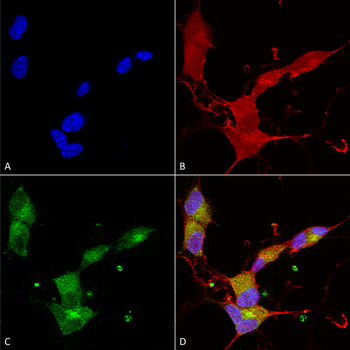
Immunocytochemistry/Immunofluorescence analysis using Mouse Anti-KCNQ4 Monoclonal Antibody, Clone N43/6. Tissue: Neuroblastoma cells (SH-SY5Y). Species: Human. Fixation: 4% PFA for 15 min. Primary Antibody: Mouse Anti-KCNQ4 Monoclonal Antibody at 1:100 for overnight at 4C with slow rocking. Secondary Antibody: AlexaFluor 488 at 1:1000 for 1 hour at RT. Counterstain: Phalloidin-iFluor 647 (red) F-Actin stain, Hoechst (blue) nuclear stain at 1:800, 1.6mM for 20 min at RT. (A) Hoechst (blue) nuclear stain. (B) Phalloidin-iFluor 647 (red) F-Actin stain. (C) KCNQ4 Antibody (D) Composite.
Immunohistochemistry analysis using Mouse Anti-KCNQ4 Monoclonal Antibody, Clone N43/6. Tissue: frozen brain section. Species: Mouse. Fixation: 10% Formalin Solution for 12-24 hours at RT. Primary Antibody: Mouse Anti-KCNQ4 Monoclonal Antibody at 1:1000 for 1 hour at RT. Secondary Antibody: HRP/DAB Detection System: Biotinylated Goat Anti-Mouse, Streptavidin Peroxidase, DAB Chromogen (brown) for 30 minutes at RT. Counterstain: Mayer Hematoxylin (purple/blue) nuclear stain at 250-500 µl for 5 minutes at RT.
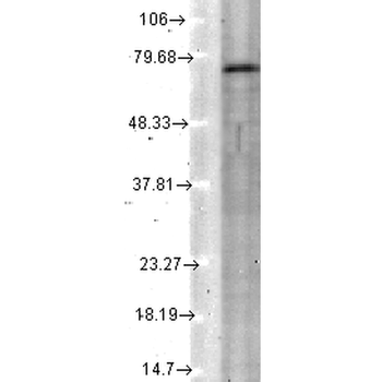
Western Blot analysis of Rat tissue lysate showing detection of KCNQ4 protein using Mouse Anti-KCNQ4 Monoclonal Antibody, Clone N43/6. Load: 15 µg. Block: 1.5% BSA for 30 minutes at RT. Primary Antibody: Mouse Anti-KCNQ4 Monoclonal Antibody at 1:1000 for 2 hours at RT. Secondary Antibody: Sheep Anti-Mouse IgG: HRP for 1 hour at RT.
Immunohistochemistry analysis using Mouse Anti-KCNQ4 Monoclonal Antibody, Clone N43/6. Tissue: hippocampus. Species: Human. Fixation: Bouins Fixative and paraffin-embedded. Primary Antibody: Mouse Anti-KCNQ4 Monoclonal Antibody at 1:1000 for 1 hour at RT. Secondary Antibody: FITC Goat Anti-Mouse (green) at 1:50 for 1 hour at RT.
* VAT and and shipping costs not included. Errors and price changes excepted