
ADRB2 Recombinant Rabbit Monoclonal Antibody, Clone: [7C1], Unconjugated
Catalog Number:
BYT-ORB1499374
- Images (9)


| Article Name: | ADRB2 Recombinant Rabbit Monoclonal Antibody, Clone: [7C1], Unconjugated |
| Biozol Catalog Number: | BYT-ORB1499374 |
| Supplier Catalog Number: | orb1499374 |
| Alternative Catalog Number: | BYT-ORB1499374-100,BYT-ORB1499374-25,BYT-ORB1499374-50 |
| Manufacturer: | Biorbyt |
| Host: | Rabbit |
| Category: | Antikörper |
| Application: | IF, IHC-Fr, IHC-P |
| Species Reactivity: | Human, Mouse, Rat |
| Immunogen: | A synthesized peptide derived from human beta 2 Adrenergic receptor (360-413/413aa) |
| Conjugation: | Unconjugated |
| Alternative Names: | beta 2-adrenergic receptor, beta 2 Adrenergic Receptor, ADRB2R, ADRBR, ADRB2_HUMAN, Adrenergic beta 2 receptor surface, B2AR, BAR, beta 2 adrenoreceptor, BETA2AR, Catecholamine receptor, beta2-adrenergic receptor. |
| ADRB2 Recombinant Rabbit Monoclonal Antibody |
| Clonality: | Recombinant |
| Concentration: | 1mg/ml |
| Clone Designation: | [7C1] |
| Molecular Weight: | 60 kDa |
| UniProt: | P07550 |
| Buffer: | 0.01M TBS (pH7.4) with 1% rAlbumin, 0.02% Proclin300 and 50% Glycerol. |
| Form: | Liquid |
| Target: | ADRB2 |
| Application Dilute: | IHC-P=1:100-500, IHC-F=1:100-500, IF=1:100-500 |
|
|
Blocking buffer: 5% NFDM/TBST, Primary Ab Dilution: 1:1000, Secondary Ab: Goat Anti-Rabbit IgG H&L (HRP), Lysate: Mouse kidney, Protein loading quantity: 20 µg, Exposure time: 60 s, Predicted MW: 46 kDa, Observed MW: 46 kDa. |
|
|
Blocking buffer: 5% NFDM/TBST, Primary Ab Dilution: 1:2000, Primary Ab incubation condition: 2 hours at room temperature, Secondary Ab: Goat Anti-Rabbit IgG H&L (HRP), Lysate: 1: A431, 2: MCF-7, 3: MDA-MB-231, 4: Rat kidney, 5: Mouse kidney, Protein loading quantity: 20 µg, Exposure time: 60 s, Predicted MW: 46 kDa, Observed MW: 46 kDa. |
|
|
Cell line: A431, Fixation: 100% Ice-cold methanol, Permeabilization: 0.1% TritonX-100, Primary Ab Dilution: 1:100, Primary Ab incubation condition: 4C overnight, Secondary Ab: Goat Anti-Rabbit IgG, Nuclear counter stain: DAPI (Blue), Comment: Color green is the positive signal for orb1499374. |
|
|
Cell line: MCF7, Fixation: 100% Ice-cold methanol, Permeabilization: 0.1% TritonX-100, Primary Ab Dilution: 1:100, Primary Ab incubation condition: 4C overnight, Secondary Ab: Goat Anti-Rabbit IgG, Nuclear counter stain: DAPI (Blue), Comment: Color green is the positive signal for orb1499374. |
|
|
Cell line: SH-SY5Y, Fixation: 100% Ice-cold methanol, Permeabilization: 0.1% TritonX-100, Primary Ab Dilution: 1:50, Primary Ab incubation condition: 4C, overnight, Secondary Ab: Goat Anti-Rabbit IgG, Nuclear counter stain: DAPI (Blue). |
|
|
Cell line: SH-SY5Y, Fixation: 4% Paraformaldehyde, Permeabilization: 90% Methanol, Primary Ab Dilution: 1:100, Secondary Ab: Goat Anti-Rabbit IgG, Unlabelled control: The cell without incubation, with primary antibody and secondary antibody, (Black line). Isotype control: Rabbit monoclonal IgG (Blue line). Comment: Line red is the positive signal for orb1499374. |
|
|
Immunohistochemical analysis of paraffin-embedded mouse brain tissue using anti-beta 2 Adrenergic Receptor antibody. The section was pre-treated using heat mediated antigen retrieval with Tris-EDTA buffer (pH 8.0-8.4) for 20 minutes. The tissues were blocked in 5% BSA for 30 minutes at room temperature, washed with ddH2O and PBS, and then probed with the primary antibody (orb1499374, 1/50) for 30 minutes at room temperature. The detection was performed using an HRP conjugated compact polymer system. DAB was used as the chromogen. Tissues were counterstained with hematoxylin and mounted with DPX. |
|
|
Immunohistochemical analysis of paraffin-embedded mouse liver tissue using anti-beta 2 Adrenergic Receptor antibody. The section was pre-treated using heat mediated antigen retrieval with Tris-EDTA buffer (pH 8.0-8.4) for 20 minutes. The tissues were blocked in 5% BSA for 30 minutes at room temperature, washed with ddH2O and PBS, and then probed with the primary antibody (orb1499374, 1/50) for 30 minutes at room temperature. The detection was performed using an HRP conjugated compact polymer system. DAB was used as the chromogen. Tissues were counterstained with hematoxylin and mounted with DPX. |
|
|
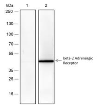
Western blot analysis of beta 2 Adrenergic Receptor on zebrafish tissue lysates. Proteins were transferred to a PVDF membrane and blocked with 5% BSA in PBS for 1 hour at room temperature. The primary antibody (orb1499374, 1/500) was used in 5% BSA at room temperature for 2 hours. Goat Anti-Rabbit IgG - HRP Secondary Antibody at 1:5000 dilution was used for 1 hour at room temperature. |
Product Guarantee and Expert Support
