136.36mM Ethanolamine, 133.23 mM Chlorides, 9.55mM Phosphates, 9.55mM Sodium Bicarbonate.
Target:
HO-1
Application Dilute:
WB (1:1000), ICC/IF (1:100)
Application Notes:
Application Notes: 1 µg/ml of SPC-112 was sufficient for detection of HO-1 in 10 µg of heat shocked HeLa cell lysate by colorimetric immunoblot analysis using Goat anti-rabbit IgG:HRP as the secondary antibody
Immunocytochemistry/Immunofluorescence analysis using Rabbit Anti-HO-1 Polyclonal Antibody. Tissue: Heat Shocked Cervical cancer cell line (HeLa). Species: Human. Fixation: 2% Formaldehyde for 20 min at RT. Primary Antibody: Rabbit Anti-HO-1 Polyclonal Antibody at 1:100 for 12 hours at 4C. Secondary Antibody: FITC Goat Anti-Rabbit (green) at 1:200 for 2 hours at RT. Counterstain: DAPI (blue) nuclear stain at 1:40000 for 2 hours at RT. Localization: Endoplasmic reticulum membrane. Cytoplasm. Magnification: 100x. Heat Shocked at 42C for 1h.
Western blot analysis of Human Cell line lysates showing detection of HO-1 protein using Rabbit Anti-HO-1 Polyclonal Antibody. Load: 15 µgprotein. Block: 1.5% BSA. Primary Antibody: Rabbit Anti-HO-1 Polyclonal Antibody at 1:1000 for 2 hours at RT. Secondary Antibody: Donkey Anti-Rabbit IgG: HRP for 1 hour at RT.
Immunocytochemistry/Immunofluorescence analysis using Rabbit Anti-HO-1 Polyclonal Antibody. Tissue: Heat Shocked Cervical cancer cell line (HeLa). Species: Human. Fixation: 2% Formaldehyde for 20 min at RT. Primary Antibody: Rabbit Anti-HO-1 Polyclonal Antibody at 1:100 for 12 hours at 4C. Secondary Antibody: APC Goat Anti-Rabbit (red) at 1:200 for 2 hours at RT. Counterstain: DAPI (blue) nuclear stain at 1:40000 for 2 hours at RT. Localization: Endoplasmic reticulum membrane. Cytoplasm. Magnification: 20x. (A) DAPI (blue) nuclear stain. (B) Anti-HO-1 Antibody. (C) Composite. Heat Shocked at 42C for 1h.
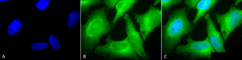
Immunocytochemistry/Immunofluorescence analysis using Rabbit Anti-HO-1 Polyclonal Antibody. Tissue: Cervical cancer cell line (HeLa). Species: Human. Fixation: 2% Formaldehyde for 20 min at RT. Primary Antibody: Rabbit Anti-HO-1 Polyclonal Antibody at 1:120 for 12 hours at 4C. Secondary Antibody: FITC Goat Anti-Rabbit (green) at 1:200 for 2 hours at RT. Counterstain: DAPI (blue) nuclear stain at 1:40000 for 2 hours at RT. Localization: Endoplasmic reticulum membrane. Cytoplasm. Magnification: 100x. (A) DAPI (blue) nuclear stain. (B) Anti-HO-1 Antibody. (C) Composite.
* VAT and and shipping costs not included. Errors and price changes excepted