Alpha B Crystallin Antibody (Biotin), Rabbit, Polyclonal
Biozol Catalog Number:
BYT-ORB151547
Supplier Catalog Number:
orb151547
Alternative Catalog Number:
BYT-ORB151547-200
Manufacturer:
Biorbyt
Host:
Rabbit
Category:
Antikörper
Application:
ICC, IF, IHC, WB
Species Reactivity:
Bovine, Gallus, Human, Mouse, Rat
Immunogen:
Synthetic peptide corresponding to human alpha B crystallin conjugated to KLH
Conjugation:
Biotin
Alternative Names:
Alpha B Crystallin, CRYAB, CRYA2, HSPB5, Heat Shock Protein Beta-5, Heat Shock Protein Family B Member 5, Alpha-Crystallin B Chain, Crystallin Alpha B, Alpha (B)-Crystallin, HEL-S-101, CMD1II, CTPP2, CTRCT16, MFM2, Rosenthal Fiber Component, Epididymis Secretory Protein Li 101, Heat-Shock 20 KD Like-Protein, Renal Carcinoma Antigen NY-REN-27
Rabbit polyclonal to Alpha B Crystallin (Biotin). The alpha-crystallins are major water-soluble lens structural proteins of the vertebrate eye that are related to the small heat shock protein family. The alpha-crystallins possess structural and functional similarities with Hsp25 and Hsp27. Mammalian lens cystallins are divided into alpha, beta and gamma families. Alpha and beta families are further divided into acidic and basic groups (Alpha-A and Alpha-B respectively). In the lens, alpha-crystallin primarily functions to maintain proper refractive index, however it can also function as a molecular chaperone that binds to the denatured proteins, keeping them in solution and thereby maintaining the translucency of the lens. When cellular stress occurs, alpha-crystallin enters its phosphorylated state and may serve a structural control function and play a role in protein maintenance. In addition to their interaction with proteins, alpha-crystallins also interact with native molecules such as membrane proteins, Golgi matrix protein, structural proteins, nuclear proteins and DNA (3, 4, 5, 6, and 7). Two other functions are an autokinase activity and participation in the intracellular architecture, and it has also been proven that both alpha-A and B prevent apoptosis by inhibiting caspases. Specifically, alpha-B cystallin is found in many cells and organs outside the lens, and alpha B is overexpressed in several neurological disorders and in cell lines under stress conditions..
136.36mM Ethanolamine, 133.23 mM Chlorides, 9.55mM Phosphates, 9.55mM Sodium Bicarbonate.
Target:
Alpha B Crystallin
Application Dilute:
WB (1:5000), ICC/IF (1:120)
Application Notes:
Application Notes: A 1:5000 dilution of SPC-126 was sufficient for detection of alpha B crystallin in 20 µg of HeLa cell lysate by ECL immunoblot analysis
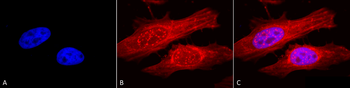
Immunocytochemistry/Immunofluorescence analysis using Rabbit Anti-Alpha B Crystallin Polyclonal Antibody. Tissue: Heat Shocked Cervical cancer cell line (HeLa). Species: Human. Fixation: 2% Formaldehyde for 20 min at RT. Primary Antibody: Rabbit Anti-Alpha B Crystallin Polyclonal Antibody at 1:120 for 12 hours at 4C. Secondary Antibody: APC Goat Anti-Rabbit (red) at 1:200 for 2 hours at RT. Counterstain: DAPI (blue) nuclear stain at 1:40000 for 2 hours at RT. Localization: Actin filament bundles. Nuclear splicing speckles. Exosomes. Magnification: 100x. (A) DAPI (blue) nuclear stain. (B) Anti-Alpha B Crystallin Antibody. (C) Composite. Heat Shocked at 42C for 1h.
Western blot analysis of Human A431, HCT116, HeLa, HepG2, HEK293, HUVEC, Jurkat, MCF7, PC3 and T98G cell lysates showing detection of ~22 kDa Alpha B Crystallin protein using Rabbit Anti-Alpha B Crystallin Polyclonal Antibody. Lane 1: Molecular Weight Ladder (MW). Lane 2: A431 cell lysates. Lane 3: HCT116 cell lysates. Lane 4: HeLa cell lysates. Lane 5: HepG2 cell lysates. Lane 6: HEK293 cell lysates. Lane 7: HUVEC cell lysates. Lane 8: Jurkat cell lysates. Lane 9: MCF7 cell lysates. Lane 10: PC3 cell lysates. Lane 11: T98G cell lysates. Load: 15 µg. Block: 5% Skim Milk in 1X TBST. Primary Antibody: Rabbit Anti-Alpha B Crystallin Polyclonal Antibody at 1:1000 for 60 min at RT. Secondary Antibody: Goat Anti-Rabbit IgG: HRP at 1:1000 for 60 min at RT. Color Development: ECL solution for 6 min in RT. Predicted/Observed Size: ~22 kDa.
Immunocytochemistry/Immunofluorescence analysis using Rabbit Anti-Alpha B Crystallin Polyclonal Antibody. Tissue: Heat Shocked Cervical cancer cell line (HeLa). Species: Human. Fixation: 2% Formaldehyde for 20 min at RT. Primary Antibody: Rabbit Anti-Alpha B Crystallin Polyclonal Antibody at 1:120 for 12 hours at 4C. Secondary Antibody: FITC Goat Anti-Rabbit (green) at 1:200 for 2 hours at RT. Counterstain: DAPI (blue) nuclear stain at 1:40000 for 2 hours at RT. Localization: Actin filament bundles. Nuclear splicing speckles. Exosomes. Magnification: 20x. (A) DAPI (blue) nuclear stain. (B) Anti-Alpha B Crystallin Antibody. (C) Composite. Heat Shocked at 42C for 1h.
Western blot analysis of Rat Brain cell lysates showing detection of ~22 kDa Alpha B Crystallin protein using Rabbit Anti-Alpha B Crystallin Polyclonal Antibody. Lane 1: Molecular Weight Ladder (MW). Lane 2: Rat Brain cell lysates. Load: 15 µg. Block: 5% Skim Milk in 1X TBST. Primary Antibody: Rabbit Anti-Alpha B Crystallin Polyclonal Antibody at 1:1000 for 60 min at RT. Secondary Antibody: Goat Anti-Rabbit IgG: HRP at 1:1000 for 60 min at RT. Color Development: ECL solution for 6 min in RT. Predicted/Observed Size: ~22 kDa.
* VAT and and shipping costs not included. Errors and price changes excepted