A synthetic peptide corresponding to a sequence at the N-terminus of human Caspase 8, different from the related mouse and rat sequences by seven amino acids.
Each vial contains antibody formulated with stabilizing components, 0.9 mg NaCl, 0.2 mg Na2HPO4, and 0.05 mg NaN3. *This antibody is supplied in a stabilized formulation. Compatibility with conjugation reactions depends on the chemistry of the conjugation
Form:
Lyophilized
Target:
Caspase-8
Application Dilute:
Western blot, 0.1-0.5 µg/ml, Human, Mouse, Rat Immunohistochemistry(Paraffin-embedded Section), 0.5-1 µg/ml, Human, Mouse, Rat Immunohistochemistry(Frozen Section), 0.5-1 µg/ml, Human Immunocytochemistry, 0.5-1 µg/ml, Human Flow Cytometry (Fixed), 1-3 µg/
Flow Cytometry analysis of Hela cells using anti-CASP8 antibody. Overlay histogram showing Hela cells (Blue line).The cells were blocked with 10% normal goat serum. And then incubated with rabbit anti-CASP8 Antibody (1 µg/1x10 6 cells) for 30 min at 20C. DyLight488 conjugated goat anti-rabbit IgG (5-10 µg/1x10 6 cells) was used as secondary antibody for 30 minutes at 20C. Isotype control antibody (Green line) was rabbit IgG (1 µg/1x10 6) used under the same conditions. Unlabelled sample (Red line) was also used as a control.
Flow Cytometry analysis of HeLa cells using anti-CASP8 antibody (Bl
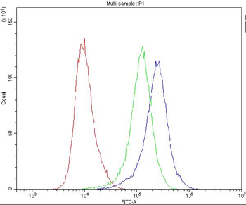
Flow Cytometry analysis of PC-3 cells using anti-CASP8 antibody. Overlay histogram showing PC-3 cells (Blue line). To facilitate intracellular staining, cells were fixed with 4% paraformaldehyde and permeabilized with permeabilization buffer. The cells were blocked with 10% normal goat serum. And then incubated with rabbit anti-CASP8 Antibody (1 µg/1x10 6 cells) for 30 min at 20C. DyLight488 conjugated goat anti-rabbit IgG (5-10 µg/1x10 6 cells) was used as secondary antibody for 30 minutes at 20C. Isotype control antibody (Green line) was rabbit IgG (1 µg/1x10 6) used under the same conditions. Unlabelled sample without incubation with primary antibody and secondary antibody (Red line) was used as a blank control.
IHC analysis of Caspase8 using anti-Caspase8 antibody. Caspase8 was detected in paraffin-embedded section of human intestinal cancer tissues. Heat mediated antigen retrieval was performed in citrate buffer (pH6, epitope retrieval solution) for 20 mins. The tissue section was blocked with 10% goat serum. The tissue section was then incubated with 1 µg/ml rabbit anti-Caspase8 Antibody overnight at 4C. Biotinylated goat anti-rabbit IgG was used as secondary antibody and incubated for 30 minutes at 37C. The tissue section was developed using Strepavidin-Biotin-Complex (SABC) with DAB as the chromogen.
IHC analysis of Caspase8 using anti-Caspase8 antibody. Caspase8 was detected in paraffin-embedded section of human mammary cancer tissues. Heat mediated antigen retrieval was performed in citrate buffer (pH6, epitope retrieval solution) for 20 mins. The tissue section was blocked with 10% goat serum. The tissue section was then incubated with 1 µg/ml rabbit anti-Caspase8 Antibody overnight at 4C. Biotinylated goat anti-rabbit IgG was used as secondary antibody and incubated for 30 minutes at 37C. The tissue section was developed using Strepavidin-Biotin-Complex (SABC) with DAB as the chromogen.
IHC analysis of Caspase8 using anti-Caspase8 antibody. Caspase8 was detected in paraffin-embedded section of mouse spleen tissues. Heat mediated antigen retrieval was performed in citrate buffer (pH6, epitope retrieval solution) for 20 mins. The tissue section was blocked with 10% goat serum. The tissue section was then incubated with 1 µg/ml rabbit anti-Caspase8 Antibody overnight at 4C. Biotinylated goat anti-rabbit IgG was used as secondary antibody and incubated for 30 minutes at 37C. The tissue section was developed using Strepavidin-Biotin-Complex (SABC) with DAB as the chromogen.
IHC analysis of Caspase8 using anti-Caspase8 antibody. Caspase8 was detected in paraffin-embedded section of rat intestine tissues. Heat mediated antigen retrieval was performed in citrate buffer (pH6, epitope retrieval solution) for 20 mins. The tissue section was blocked with 10% goat serum. The tissue section was then incubated with 1 µg/ml rabbit anti-Caspase8 Antibody overnight at 4C. Biotinylated goat anti-rabbit IgG was used as secondary antibody and incubated for 30 minutes at 37C. The tissue section was developed using Strepavidin-Biotin-Complex (SABC) with DAB as the chromogen.
IHC analysis of Caspase8 using anti-Caspase8 antibody. Caspase8 was detected in paraffin-embedded section of rat spleen tissues. Heat mediated antigen retrieval was performed in citrate buffer (pH6, epitope retrieval solution) for 20 mins. The tissue section was blocked with 10% goat serum. The tissue section was then incubated wit
* VAT and and shipping costs not included. Errors and price changes excepted