
CLOCK Recombinant Rabbit Monoclonal Antibody, Clone: [3F9], Unconjugated
Catalog Number:
BYT-ORB1816868
- Images (8)


| Article Name: | CLOCK Recombinant Rabbit Monoclonal Antibody, Clone: [3F9], Unconjugated |
| Biozol Catalog Number: | BYT-ORB1816868 |
| Supplier Catalog Number: | orb1816868 |
| Alternative Catalog Number: | BYT-ORB1816868-100,BYT-ORB1816868-50 |
| Manufacturer: | Biorbyt |
| Host: | Rabbit |
| Category: | Antikörper |
| Application: | FC, ICC, IF, IHC-Fr, IHC-P |
| Species Reactivity: | Human, Mouse, Rat |
| Immunogen: | A synthesized peptide derived from human CLOCK (145-180aa) |
| Conjugation: | Unconjugated |
| Alternative Names: | KAT13D, bHLHe8, 5330400M04Rik, CLOCK_HUMAN, CLOCK, hCLOCK, Class E basic helix-loop-helix protein 8 (bHLHe8), 2.3.1.48, KIAA0334, CLOCK_MOUSE, mCLOCK, CLOCK_RAT, rCLOCK, |
| CLOCK Recombinant Rabbit Monoclonal Antibody |
| Clonality: | Recombinant |
| Concentration: | 1mg/ml |
| Clone Designation: | [3F9] |
| Molecular Weight: | 95 kDa |
| UniProt: | O15516 |
| Buffer: | 1*TBS (pH7.4), 0.05% rAlbumin, 40% Glycerol. Preservative: 0.02% Proclin300. |
| Form: | Liquid |
| Target: | CLOCK |
| Application Dilute: | IHC-P=1:100-500, IHC-F=1:100-500, ICC/IF=1:50-200, IF=1:100-500, Flow-Cyt=1:50-100 |
|
|
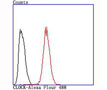
Flow cytometric analysis of CLOCK was done on Hela cells. The cells were fixed, permeabilized and stained with the primary antibody (orb1816868, 1/50) (red). After incubation of the primary antibody at room temperature for an hour, the cells were stained with a Alexa Fluor488 conjugate-Goat anti-Rabbit IgG Secondary antibody at 1/1000 dilution for 30 minutes. Unlabelled sample was used as a control (cells without incubation with primary antibody, black). |
|
|
ICC staining of CLOCK in Hela cells (green). Formalin fixed cells were permeabilized with 0.1% Triton X-100 in TBS for 10 minutes at room temperature and blocked with 10% negative goat serum for 15 minutes at room temperature. Cells were probed with the primary antibody (orb1816868, 1/50) for 1 hour at room temperature, washed with PBS. Alexa Fluor488 conjugate-Goat anti-Rabbit IgG was used as the secondary antibody at 1/1000 dilution. The nuclear counter stain is DAPI (blue). |
|
|
ICC staining of CLOCK in SHG-44 cells (green). Formalin fixed cells were permeabilized with 0.1% Triton X-100 in TBS for 10 minutes at room temperature and blocked with 10% negative goat serum for 15 minutes at room temperature. Cells were probed with the primary antibody (orb1816868, 1/50) for 1 hour at room temperature, washed with PBS. Alexa Fluor488 conjugate-Goat anti-Rabbit IgG was used as the secondary antibody at 1/1000 dilution. The nuclear counter stain is DAPI (blue). |
|
|
Paraformaldehyde-fixed, paraffin embedded (human colon carcinoma), Antigen retrieval by boiling in sodium citrate buffer (pH6.0) for 15 min, Block endogenous peroxidase by 3% hydrogen peroxide for 20 minutes, Blocking buffer (normal goat serum) at 37C for 30 min, Antibody incubation with (CLOCK) Monoclonal Antibody, Unconjugated (orb1816868) at 1:200 overnight at 4C, followed by operating according to SP Kit (Rabbit) instructionsand DAB staining. |
|
|
Paraformaldehyde-fixed, paraffin embedded (human colon), Antigen retrieval by boiling in sodium citrate buffer (pH6.0) for 15 min, Block endogenous peroxidase by 3% hydrogen peroxide for 20 minutes, Blocking buffer (normal goat serum) at 37C for 30 min, Antibody incubation with (CLOCK) Monoclonal Antibody, Unconjugated (orb1816868) at 1:200 overnight at 4C, followed by operating according to SP Kit (Rabbit) instructionsand DAB staining. |
|
|
Paraformaldehyde-fixed, paraffin embedded (human fetal skeletal tissue), Antigen retrieval by boiling in sodium citrate buffer (pH6.0) for 15 min, Block endogenous peroxidase by 3% hydrogen peroxide for 20 minutes, Blocking buffer (normal goat serum) at 37C for 30 min, Antibody incubation with (CLOCK) Monoclonal Antibody, Unconjugated (orb1816868) at 1:200 overnight at 4C, followed by operating according to SP Kit (Rabbit) instructionsand DAB staining. |
|
|
Paraformaldehyde-fixed, paraffin embedded (human kidney), Antigen retrieval by boiling in sodium citrate buffer (pH6.0) for 15 min, Block endogenous peroxidase by 3% hydrogen peroxide for 20 minutes, Blocking buffer (normal goat serum) at 37C for 30 min, Antibody incubation with (CLOCK) Monoclonal Antibody, Unconjugated (orb1816868) at 1:200 overnight at 4C, followed by operating according to SP Kit (Rabbit) instructionsand DAB staining. |
|
|
Western blot analysis of CLOCK on different lysates. Proteins were transferred to a PVDF membrane and blocked with 5% BSA in PBS for 1 hour at room temperature. The primary antibody (1/500) was used in 5% BSA at room temperature for 2 hours. Goat Anti-Rabbit IgG - HRP Secondary Antibody at 1:200000 dilution was used for 1 hour at room temperature. Positive control: Lane 1: MCF-7 cell lysate, Lane 2: PC-12 cell lysate. |
Product Guarantee and Expert Support
