
BCAT2 Antibody, Unconjugated, Rabbit, Polyclonal
Catalog Number:
BYT-ORB1819416
- Images (8)


| Article Name: | BCAT2 Antibody, Unconjugated, Rabbit, Polyclonal |
| Biozol Catalog Number: | BYT-ORB1819416 |
| Supplier Catalog Number: | orb1819416 |
| Alternative Catalog Number: | BYT-ORB1819416-100 |
| Manufacturer: | Biorbyt |
| Host: | Rabbit |
| Category: | Antikörper |
| Application: | ELISA, FC, IF, IHC, WB |
| Species Reactivity: | Human, Mouse, Rat |
| Immunogen: | E.coli-derived human BCAT2 recombinant protein (Position: A11-E363). Human BCAT2 shares 83.6% and 81.9% amino acid (aa) sequence identity with mouse and rat BCAT2, respectively. |
| Conjugation: | Unconjugated |
| Alternative Names: | 70 kDa ribosomal protein S6 kinase 1 antibody, KS6B1_HUMAN antibody, p70 alpha antibody, P70 beta 1 antibody, p70 ribosomal S6 kinase alpha antibody, p70 ribosomal S6 kinase beta 1 antibody, p70 S6 kinase alpha antibody, P70 S6 Kinase antibody, p70 S6 kinase alpha 1 antibody, p70 S6 kinase alpha 2 antibody, p70 S6K antibody, p70 S6K-alpha antibody, p70 S6KA antibody, p70(S6K) alpha antibody, p70(S6K)-alpha antibody, p70-alpha antibody, p70-S6K 1 antibody, p70-S6K antibody, P70S6K antibody, P70S6K1 antibody, p70S6Kb antibody, PS6K antibody, Ribosomal protein S6 kinase 70kDa polypeptide 1 antibody, Ribosomal protein S6 kinase beta 1 antibody, Ribosomal protein S6 kinase beta-1 antibody, Ribosomal protein S6 kinase I antibody, RPS6KB1 antibody, S6K antibody, S6K-beta-1 antibody, S6K1 antibody, Serine/threonine kinase 14 alpha antibody, Serine/threonine-protein kinase 14A antibody, STK14A antibody |
| Anti-BCAT2 Antibody. Tested in ELISA, IF, IHC, WB, Flow Cytometry applications. This antibody reacts with Human, Mouse, Rat. |
| Clonality: | Polyclonal |
| Concentration: | Adding 0.2 ml of distilled water will yield a concentration of 500 µg/ml. |
| Molecular Weight: | 39 kDa |
| UniProt: | O15382 |
| Form: | Lyophilized |
| Target: | Serine/threonine-protein kinase AtPK1/AtPK6 |
| Application Notes: | Application Notes: Western blot, 0.25-0.5 µg/ml, Human, Mouse, Rat Immunohistochemistry, 2-5 µg/ml, Human Immunofluorescence, 5 µg/ml, Human Flow Cytometry (Fixed), 1-3 µg/1x106 cells, Human ELISA, 0.1-0.5 µg/ml, -. Adding 0.2 ml of distilled water will yield a concentration of 500 µg/ml |
|
|
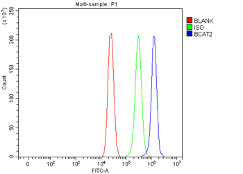
Flow Cytometry analysis of MCF-7 cells using anti-BCAT2 antibody. Overlay histogram showing MCF-7 cells (Blue line). To facilitate intracellular staining, cells were fixed with 4% paraformaldehyde and permeabilized with permeabilization buffer. The cells were blocked with 10% normal goat serum. And then incubated with rabbit anti-BCAT2 Antibody (1 µg/1x10 6 cells) for 30 min at 20C. DyLight488 conjugated goat anti-rabbit IgG (5-10 µg/1x10 6 cells) was used as secondary antibody for 30 minutes at 20C. Isotype control antibody (Green line) was rabbit IgG (1 µg/1x10 6) used under the same conditions. Unlabelled sample (Red line) was also used as a control. |
|
|
IF analysis of BCAT2 using anti-BCAT2 antibody. BCAT2 was detected in a paraffin-embedded section of human intestinal cancer tissue. Heat mediated antigen retrieval was performed in EDTA buffer (pH8.0, epitope retrieval solution). The tissue section was blocked with 10% goat serum. The tissue section was then incubated with 5 µg/mL rabbit anti-BCAT2 Antibody overnight at 4C. Cy3 Conjugated Goat Anti-Rabbit IgG was used as secondary antibody at 1:500 dilution and incubated for 30 minutes at 37C. The section was counterstained with DAPI. Visualize using a fluorescence microscope and filter sets appropriate for the label used. |
|
|
IHC analysis of BCAT2 using anti-BCAT2 antibody. BCAT2 was detected in a paraffin-embedded section of human colon adenocarcinoma tissue. Heat mediated antigen retrieval was performed in EDTA buffer (pH8.0, epitope retrieval solution). The tissue section was blocked with 10% goat serum. The tissue section was then incubated with 2 µg/ml rabbit anti-BCAT2 Antibody overnight at 4C. Peroxidase Conjugated Goat Anti-rabbit IgG was used as secondary antibody and incubated for 30 minutes at 37C. The tissue section was developed using HRP Conjugated Rabbit IgG Super Vision Assay Kit with DAB as the chromogen. |
|
|
IHC analysis of BCAT2 using anti-BCAT2 antibody. BCAT2 was detected in a paraffin-embedded section of human liver cancer tissue. Heat mediated antigen retrieval was performed in EDTA buffer (pH8.0, epitope retrieval solution). The tissue section was blocked with 10% goat serum. The tissue section was then incubated with 2 µg/ml rabbit anti-BCAT2 Antibody overnight at 4C. Peroxidase Conjugated Goat Anti-rabbit IgG was used as secondary antibody and incubated for 30 minutes at 37C. The tissue section was developed using HRP Conjugated Rabbit IgG Super Vision Assay Kit with DAB as the chromogen. |
|
|
IHC analysis of BCAT2 using anti-BCAT2 antibody. BCAT2 was detected in a paraffin-embedded section of human lung cancer tissue. Heat mediated antigen retrieval was performed in EDTA buffer (pH8.0, epitope retrieval solution). The tissue section was blocked with 10% goat serum. The tissue section was then incubated with 2 µg/ml rabbit anti-BCAT2 Antibody overnight at 4C. Peroxidase Conjugated Goat Anti-rabbit IgG was used as secondary antibody and incubated for 30 minutes at 37C. The tissue section was developed using HRP Conjugated Rabbit IgG Super Vision Assay Kit with DAB as the chromogen. |
|
|
IHC analysis of BCAT2 using anti-BCAT2 antibody. BCAT2 was detected in a paraffin-embedded section of human urothelial carcinoma tissue. Heat mediated antigen retrieval was performed in EDTA buffer (pH8.0, epitope retrieval solution). The tissue section was blocked with 10% goat serum. The tissue section was then incubated with 2 µg/ml rabbit anti-BCAT2 Antibody overnight at 4C. Peroxidase Conjugated Goat Anti-rabbit IgG was used as secondary antibody and incubated for 30 minutes at 37C. The tissue section was developed using HRP Conjugated Rabbit IgG Super Vision Assay Kit with DAB as the chromogen. |
|
|
Western blot analysis of BCAT2 using anti-BCAT2 antibody. Electrophoresis was performed on a 5-20% SDS-PAGE gel at 70V (Stacking gel) / 90V (Resolving gel) for 2-3 hours. The sample well of each lane was loaded with 30 ug of sample under reducing conditions. Lane 1: human RT4 |
|
|
Product Guarantee and Expert Support
