136.36mM Ethanolamine, 133.23 mM Chlorides, 9.55mM Phosphates, 9.55mM Sodium Bicarbonate
Target:
GABA A Receptor
Application Dilute:
WB (1:1000), IHC (1:1000), ICC/IF (1:100)
Application Notes:
Application Notes: 1 µg/ml of SMC-339 was sufficient for detection of Beta 3 GABA receptor in 10 µg of rat brain lysate by colorimetric immunoblot analysis using goat anti-mouse IgG:HRP as the secondary antibody
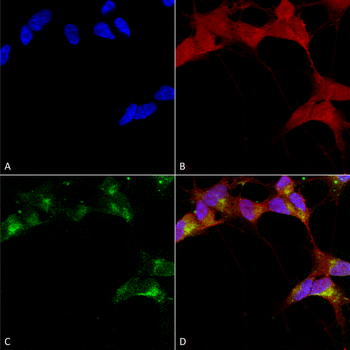
Immunocytochemistry/Immunofluorescence analysis using Mouse Anti-GABA-A Receptor Beta 3 Monoclonal Antibody, Clone N87/25. Tissue: Neuroblastoma cells (SH-SY5Y). Species: Human. Fixation: 4% PFA for 15 min. Primary Antibody: Mouse Anti-GABA-A Receptor Beta 3 Monoclonal Antibody at 1:100 for overnight at 4C with slow rocking. Secondary Antibody: AlexaFluor 488 at 1:1000 for 1 hour at RT. Counterstain: Phalloidin-iFluor 647 (red) F-Actin stain, Hoechst (blue) nuclear stain at 1:800, 1.6mM for 20 min at RT. (A) Hoechst (blue) nuclear stain. (B) Phalloidin-iFluor 647 (red) F-Actin stain. (C) GABA-A Receptor Beta 3 Antibody (D) Composite.
Immunohistochemistry analysis using Mouse Anti-GABA A Receptor Monoclonal Antibody, Clone N87/25. Tissue: Brain. Species: mouse. Fixation: 10% Formalin Solution for 12-24 hours at RT. Primary Antibody: Mouse Anti-GABA A Receptor Monoclonal Antibody at 1:1000 for 1 hour at RT. Secondary Antibody: HRP/DAB Detection System: Biotinylated Goat Anti-Mouse, Streptavidin Peroxidase, DAB Chromogen (brown) for 30 minutes at RT. Counterstain: Mayer Hematoxylin (purple/blue) nuclear stain at 250-500 µl for 5 minutes at RT.
Western Blot analysis of Rat brain membrane lysate showing detection of GABA A Receptor protein using Mouse Anti-GABA A Receptor Monoclonal Antibody, Clone N87/25. Load: 15 µg. Block: 1.5% BSA for 30 minutes at RT. Primary Antibody: Mouse Anti-GABA A Receptor Monoclonal Antibody at 1:1000 for 2 hours at RT. Secondary Antibody: Sheep Anti-Mouse IgG: HRP for 1 hour at RT.
* VAT and and shipping costs not included. Errors and price changes excepted