anti NOS1 antibody, anti nitric oxide synthase 1 (neuronal) antibody, anti NOS antibody, anti neuronal nitric oxide synthase antibody, anti PnNOS antibody, anti penile neuronal nitric oxide synthase antibody, anti penile neuronal NOS antibody, anti IHPS1 antibody, anti nNOS antibody, anti nitric oxide synthase 1, neuronal antibody
Goat polyclonal antibody to NOS1
Clonality:
Polyclonal
Molecular Weight:
161, 164.6, 125
Buffer:
Supplied at 0.5 mg/ml in Tris saline, 0.02% sodium azide, pH 7.3 with 0.5% bovine serum albumin. Aliquot and store at -20C. Minimize freezing and thawing.
Application Notes: ELISA: Peptide ELISA: antibody detection limit dilution 1:64000.IHC: This product has been sucessfully used for IHC on Olfactory bulb in mice (PMID: 20140458)WB: Approx 160-170kDa band observed in Human Skeletal Muscle and Mouse Brain lysates (calculated MW of 161kDa according to NP_000611.1). Recommended concentration: 0.3-1 µg/ml.Experiment Notes: Immunofluorescence: This product has been successfully used for IF as reported (PMID: 20140458)
1 µg/mL staining of Mouse Brain lysate (35 µg protein in RIPA buffer). Detected by chemiluminescence.
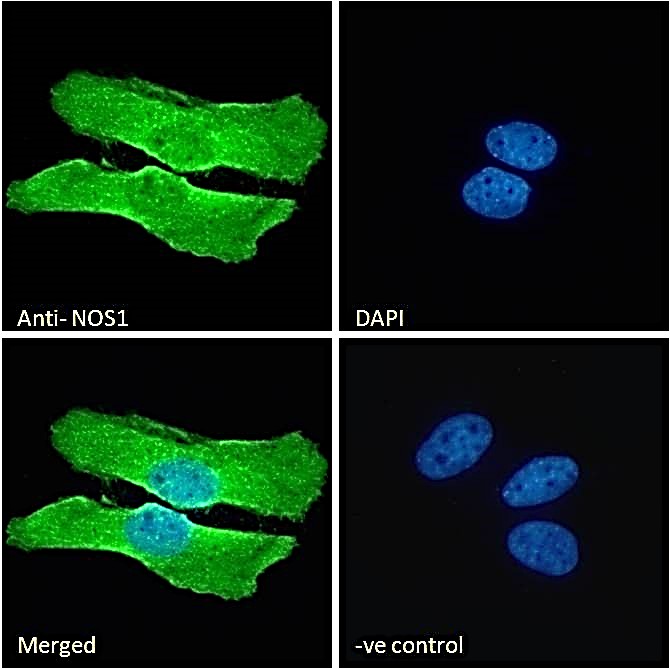
Immunofluorescence analysis of paraformaldehyde fixed HeLa cells, permeabilized with 0.15% Triton. Primary incubation 1 hr (10 µg/mL) followed by Alexa Fluor 488 secondary antibody (2 µg/mL), showing nuclear staining. The nuclear stain is DAPI (blue). Negative control: Unimmunized goat IgG (10 µg/mL) followed by Alexa Fluor 488 secondary antibody (2 µg/mL).
Immunofluorescence analysis of paraformaldehyde fixed U2OS cells, permeabilized with 0.15% Triton. Primary incubation 1 hr (10 µg/mL) followed by Alexa Fluor 488 secondary antibody (2 µg/mL), showing nuclear staining. The nuclear stain is DAPI (blue). Negative control: Unimmunized goat IgG (10 µg/mL) followed by Alexa Fluor 488 secondary antibody (2 µg/mL).
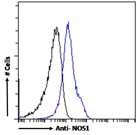
Flow cytometric analysis of paraformaldehyde fixed HeLa cells (blue line), permeabilized with 0.5% Triton. Primary incubation 1 hr (10 µg/mL) followed by Alexa Fluor 488 secondary antibody (1 µg/mL). IgG control: Unimmunized goat IgG (black line) followed by Alexa Fluor 488 secondary antibody.
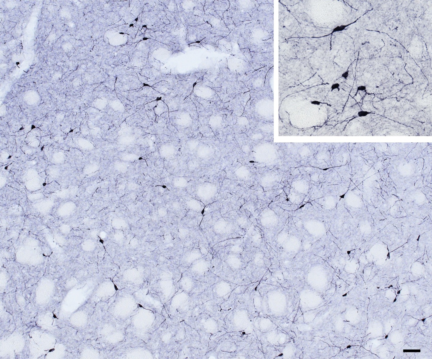
Immunostaining of 30 µm thick cryosections of PFA-perfused Human Hypothalamus, antigen retrieval with citrate buffer Ph 6 at 80C for 30 min, HRP-staining with Ni-DAB after Biotin-SP-antigoat amplification.
Immunohistochemical staining of mouse caudate-putamen using NOS1 antibody
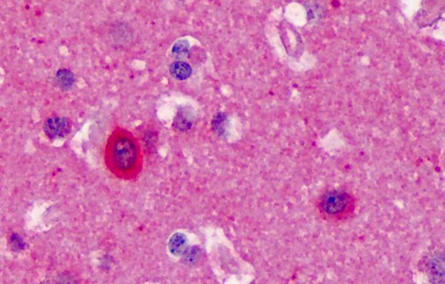
Immunohistochemical staining of Human Cortex using NOS1 antibody
Immunofluorescence analysis of HeLa cells using NOS1 antibody
Flow cytometric analysis of Kelly cells using NOS1 antibody
* VAT and and shipping costs not included. Errors and price changes excepted