STAT3 (isoform 1 and 2) Antibody, Unconjugated, Goat, Polyclonal
Biozol Catalog Number:
BYT-ORB18540
Supplier Catalog Number:
orb18540
Alternative Catalog Number:
BYT-ORB18540-100
Manufacturer:
Biorbyt
Host:
Goat
Category:
Antikörper
Application:
ELISA, FC, IF, IHC
Species Reactivity:
Human, Mouse
Conjugation:
Unconjugated
Alternative Names:
anti STAT3 antibody, anti signal transducer and activator of transcription 3 (acute-phase response factor) antibody, anti APRF antibody, anti DNA-binding protein APRF antibody, anti acute-phase response factor antibody, anti FLJ20882 antibody, anti MGC16063 antibody, anti HIES antibody
Goat polyclonal antibody to STAT3
Clonality:
Polyclonal
Concentration:
4-8 µg
Molecular Weight:
88.0, 88.1
Buffer:
Supplied at 0.5 mg/ml in Tris saline, 0.02% sodium azide, pH 7.3 with 0.5% bovine serum albumin. Aliquot and store at -20C. Minimize freezing and thawing.
Sequence:
DMELTSECATSPM
Target:
STAT3 (isoform 1 and 2)
Application Dilute:
ELISA: 1:8000, WB: 2 µg/ml, IHC-P: 4-6 µg/ml
Application Notes:
Application Notes: ELISA: Peptide ELISA: antibody detection limit dilution 1:8000.IE: An anonymous customer found positive results in IF on Human HEK 293 cells. IF, An anonymous customer found positive results in IF on Rat glial processes and nuclei.IHC: In paraffin embedded Mouse Thymus shows preferential staining of the medula over the cortex. Recommended concentration, 4-8µg/ml.WB: Approx. 90kDa band observed in lysates of NIH3T3 (calculated MW of 88.0kDa according to NP_644805.1 and of 88.1 according to NP_003141.2). Recommended concentration: 1-3 µg/ml
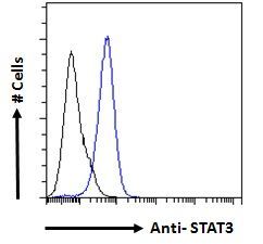
Flow Cytometry analysis of A431 cells of STATE3 antibody
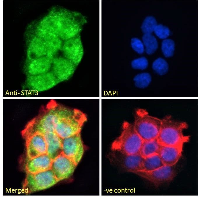
Immunofluorescence analysis of A431 cells of STAT3 antibody
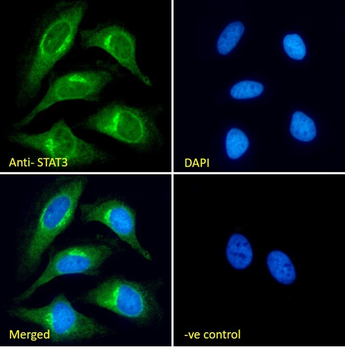
Immunofluorescence analysis of HeLa cells of STAT3 antibody
Immunofluorescence analysis of paraformaldehyde fixed HeLa cells, permeabilized with 0.15% Triton. Primary incubation 1hr (10 ug/ml) followed by Alexa Fluor 488 secondary antibody (2 ug/ml), showing cytoplasmic and Golgi apparatus staining. The nuclear stain is DAPI (blue). Negative control: Unimmunized goat IgG (10 ug/ml) followed by Alexa Fluor 488 secondary antibody (2 ug/ml).
Immunofluorescence analysis of paraformaldehyde fixed A431 cells, permeabilized with 0.15% Triton. Primary incubation 1hr (10 ug/ml) followed by Alexa Fluor 488 secondary antibody (2 ug/ml), showing cytoplasmic and staining. The nuclear stain is DAPI (blue). Negative control: Unimmunized goat IgG (10 ug/ml) followed by Alexa Fluor 488 secondary antibody (2 ug/ml).
Flow cytometric analysis of paraformaldehyde fixed A431 cells (blue line), permeabilized with 0.5% Triton. Primary incubation 1hr (10 ug/ml) followed by Alexa Fluor 488 secondary antibody (1 ug/ml). IgG control: Unimmunized goat IgG (black line) followed by Alexa Fluor 488 secondary antibody.
7 µg/ml staining of paraffin embedded Human Liver. Heat induced antigen retrieval with citrate buffer pH6, HRP-staining.
Negative Control showing staining of paraffin embedded Human Liver, with no primary antibody.
* VAT and and shipping costs not included. Errors and price changes excepted