E.coli-derived human HSPB8/Hsp22 recombinant protein (Position: M1-T196). Human HSPB8/Hsp22 shares 94.4% and 95.4% amino acid (aa) sequence identity with mouse and rat HSPB8/Hsp22, respectively.
Conjugation:
Unconjugated
Alternative Names:
Heat shock protein beta-8, HspB8, Alpha-crystallin C chain, E2-induced gene 1 protein, Protein kinase H11, Small stress protein-like protein HSP22, HSPB8, CRYAC, E2IG1, HSP22, PP1629
HSPB8/Hsp22 Rabbit Polyclonal Antibody
Clonality:
Polyclonal
Concentration:
Adding 0.2 ml of distilled water will yield a concentration of 500 µg/ml.
Western blot, 0.1-0.5µg/ml, Human, Mouse, Rat Immunohistochemistry (Paraffin-embedded Section), 0.5-1µg/ml, Human, Mouse, Rat Immunocytochemistry/Immunofluorescence, 2µg/ml, Human Immunoprecipitation, 0.5-2 µg/ml, Human Flow Cytometry (Fixed), 1-3µg/1x10
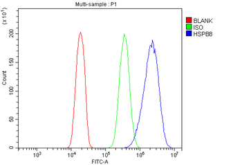
Flow Cytometry analysis of U2OS cells using anti-HSPB8/Hsp22 antibody. Overlay histogram showing U2OS cells (Blue line). To facilitate intracellular staining, cells were fixed with 4% paraformaldehyde and permeabilized with permeabilization buffer. The cells were blocked with 10% normal goat serum. And then incubated with rabbit anti-HSPB8/Hsp22 Antibody (1 µg/1x10 6 cells) for 30 min at 20C. DyLight488 conjugated goat anti-rabbit IgG (5-10 µg/1x10 6 cells) was used as secondary antibody for 30 minutes at 20C. Isotype control antibody (Green line) was rabbit IgG (1 µg/1x10 6) used under the same conditions. Unlabelled sample without incubation with primary antibody and secondary antibody (Red line) was used as a blank control.
WB analysis of HSPB8/Hsp22 using anti-HSPB8/Hsp22 antibody.Lan
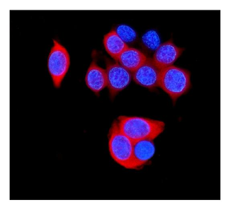
IF analysis of HSPB8/Hsp22 using anti-HSPB8/Hsp22 antibody. HSPB8/Hsp22 was detected in immunocytochemical section of MCF7 cells. Enzyme antigen retrieval was performed using IHC enzyme antigen retrieval reagent for 15 mins. The cells were blocked with 10% goat serum. And then incubated with 2 µg/mL rabbit anti-HSPB8/Hsp22 Antibody overnight at 4C. DyLight594 Conjugated Goat Anti-Rabbit IgG was used as secondary antibody at 1:100 dilution and incubated for 30 minutes at 37C. The section was counterstained with DAPI. Visualize using a fluorescence microscope and filter sets appropriate for the label used.
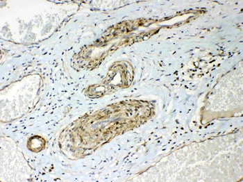
IHC analysis of HSPB8/Hsp22 using anti-HSPB8/Hsp22 antibody. HSPB8/Hsp22 was detected in paraffin-embedded section of human lung cancer tissues. Heat mediated antigen retrieval was performed in citrate buffer (pH6, epitope retrieval solution) for 20 mins. The tissue section was blocked with 10% goat serum. The tissue section was then incubated with 1 µg/ml rabbit anti-HSPB8/Hsp22 Antibody overnight at 4C. Biotinylated goat anti-rabbit IgG was used as secondary antibody and incubated for 30 minutes at 37C. The tissue section was developed using Strepavidin-Biotin-Complex (SABC) with DAB as the chromogen.
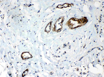
IHC analysis of HSPB8/Hsp22 using anti-HSPB8/Hsp22 antibody. HSPB8/Hsp22 was detected in paraffin-embedded section of human mammary cancer tissues. Heat mediated antigen retrieval was performed in citrate buffer (pH6, epitope retrieval solution) for 20 mins. The tissue section was blocked with 10% goat serum. The tissue section was then incubated with 1 µg/ml rabbit anti-HSPB8/Hsp22 Antibody overnight at 4C. Biotinylated goat anti-rabbit IgG was used as secondary antibody and incubated for 30 minutes at 37C. The tissue section was developed using Strepavidin-Biotin-Complex (SABC) with DAB as the chromogen.
IHC analysis of HSPB8/Hsp22 using anti-HSPB8/Hsp22 antibody. HSPB8/Hsp22 was detected in paraffin-embedded section of mouse pancreas tissues. Heat mediated antigen retrieval was performed in citrate buffer (pH6, epitope retrieval solution) for 20 mins. The tissue section was blocked with 10% goat serum. The tissue section was then incubated with 1 µg/ml rabbit anti-HSPB8/Hsp22 Antibody overnight at 4C. Biotinylated goat anti-rabbit IgG was used as secondary antibody and incubated for 30 minutes at 37C. The tissue section was developed using Strepavidin-Biotin-Complex (SABC) with DAB as the chromogen.
IHC analysis of HSPB8/Hsp22 using anti-HSPB8/Hsp22 antibody. HSPB8/Hsp22 was detected in paraffin-embedded section of rat pancreas tissues. Heat mediated antigen retrieval was performed in citrate buffer (pH6, epitope retrieval solution) for 20 mins. The tissue section was blocked with 10% goat serum. The tissue section was then incubated with 1 µg/ml rabbit anti-HSPB8/Hsp22 Antibody overnight at 4C. Biotinylated goat anti-rabbit IgG was used as secondary antibody and incubated for 30 minutes at 37C. The tissue section was developed using Strepavidin-Biotin-Complex (SABC) with DAB as the chromogen.
Immunoprecipitating HSPB8/Hsp22 in MCF-7 whole cell lysate. Western blot analysis of HSPB8/Hsp22 using anti-HSPB8/Hsp22 antibody, Lane 1: MCF-7 whole cell lysates (30 ug), Lane 2: Rabbit control Ig
* VAT and and shipping costs not included. Errors and price changes excepted