E.coli-derived human CPI17 alpha recombinant protein (Position: L30-Q126). Human CPI17 alpha shares 83.5% and 84.5% amino acid (aa) sequence identity with mouse and rat CPI17 alpha, respectively.
Conjugation:
Unconjugated
Alternative Names:
Protein phosphatase 1 regulatory subunit 14A, 17 kDa PKC-potentiated inhibitory protein of PP1, Protein kinase C-potentiated inhibitor protein of 17 kDa, CPI-17, PPP1R14A, CPI17, PPP1INL
CPI17 alpha/PPP1R14A Rabbit Polyclonal Antibody
Clonality:
Polyclonal
Concentration:
Adding 0.2 ml of distilled water will yield a concentration of 500 µg/ml.
Western blot, 0.1-0.5µg/ml, Human, Mouse, Rat Immunohistochemistry (Paraffin-embedded Section), 0.5-1µg/ml, Human, Mouse, Rat Immunocytochemistry/Immunofluorescence, 2µg/ml, Human Flow Cytometry (Fixed), 1-3µg/1x10 6 cells, Human
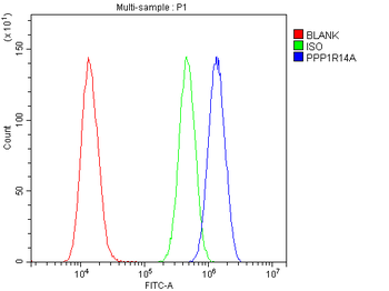
Flow Cytometry analysis of U20S cells using anti-CPI17 alpha antibody. Overlay histogram showing U20S cells (Blue line). To facilitate intracellular staining, cells were fixed with 4% paraformaldehyde and permeabilized with permeabilization buffer. The cells were blocked with 10% normal goat serum. And then incubated with rabbit anti-CPI17 alpha Antibody (1 µg/1x10 6 cells) for 30 min at 20C. DyLight488 conjugated goat anti-rabbit IgG (5-10 µg/1x10 6 cells) was used as secondary antibody for 30 minutes at 20C. Isotype control antibody (Green line) was rabbit IgG (1 µg/1x10 6) used under the same conditions. Unlabelled sample (Red line) was also used as a control.
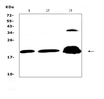
WB analysis of CPI17 alpha using anti-CPI17 alpha antibody.La
IF analysis of CPI17 alpha using anti-CPI17 alpha antibody. CPI17 alpha was detected in immunocytochemical section of U20S cells. Enzyme antigen retrieval was performed using IHC enzyme antigen retrieval reagent for 15 mins. The cells were blocked with 10% goat serum. And then incubated with 2 µg/mL rabbit anti-CPI17 alpha Antibody overnight at 4C. DyLight488 Conjugated Goat Anti-Rabbit IgG was used as secondary antibody at 1:100 dilution and incubated for 30 minutes at 37C. The section was counterstained with DAPI. Visualize using a fluorescence microscope and filter sets appropriate for the label used.
IHC analysis of CPI17 alpha using anti-CPI17 alpha antibody. CPI17 alpha was detected in paraffin-embedded section of human intestinal cancer tissues. Heat mediated antigen retrieval was performed in citrate buffer (pH6, epitope retrieval solution) for 20 mins. The tissue section was blocked with 10% goat serum. The tissue section was then incubated with 1 µg/ml rabbit anti-CPI17 alpha Antibody overnight at 4C. Biotinylated goat anti-rabbit IgG was used as secondary antibody and incubated for 30 minutes at 37C. The tissue section was developed using Strepavidin-Biotin-Complex (SABC) with DAB as the chromogen.
IHC analysis of CPI17 alpha using anti-CPI17 alpha antibody. CPI17 alpha was detected in paraffin-embedded section of human lung cancer tissues. Heat mediated antigen retrieval was performed in citrate buffer (pH6, epitope retrieval solution) for 20 mins. The tissue section was blocked with 10% goat serum. The tissue section was then incubated with 1 µg/ml rabbit anti-CPI17 alpha Antibody overnight at 4C. Biotinylated goat anti-rabbit IgG was used as secondary antibody and incubated for 30 minutes at 37C. The tissue section was developed using Strepavidin-Biotin-Complex (SABC) with DAB as the chromogen.
IHC analysis of CPI17 alpha using anti-CPI17 alpha antibody. CPI17 alpha was detected in paraffin-embedded section of human placenta tissues. Heat mediated antigen retrieval was performed in citrate buffer (pH6, epitope retrieval solution) for 20 mins. The tissue section was blocked with 10% goat serum. The tissue section was then incubated with 1 µg/ml rabbit anti-CPI17 alpha Antibody overnight at 4C. Biotinylated goat anti-rabbit IgG was used as secondary antibody and incubated for 30 minutes at 37C. The tissue section was developed using Strepavidin-Biotin-Complex (SABC) with DAB as the chromogen.
IHC analysis of CPI17 alpha using anti-CPI17 alpha antibody. CPI17 alpha was detected in paraffin-embedded section of mouse testis tissues. Heat mediated antigen retrieval was performed in citrate buffer (pH6, epitope retrieval solution) for 20 mins. The tissue section was blocked with 10% goat serum. The tissue section was then incubated with 1 µg/ml rabbit anti-CPI17 alpha Antibody overnight at 4C. Biotinylated goat anti-rabbit IgG was used as secondary antibody and incubated for 30 minutes at 37C. The tissue section was developed using Strepavidin-Biotin-Complex (SABC) with DAB as the chromogen.
IHC analysis of CPI17 alpha using anti-CPI17 alpha antibody. CPI17 alpha was detected in paraffin-embedded section of rat lung tissues. Heat mediated antigen retrieval was performed in citrate buffer (pH6, epitope retrieval solution) for 20 mins. The tissue s
* VAT and and shipping costs not included. Errors and price changes excepted