anti NHEJ1 antibody, anti XRCC4-like factor antibody, anti nonhomologous end-joining factor 1 antibody, anti FLJ12610 antibody, anti XLF antibody, anti Cernunnos antibody
Goat polyclonal antibody to NHEJ1
Clonality:
Polyclonal
Molecular Weight:
33.3
Buffer:
Supplied at 0.5 mg/ml in Tris saline, 0.02% sodium azide, pH 7.3 with 0.5% bovine serum albumin. Aliquot and store at -20C. Minimize freezing and thawing.
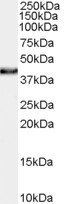
Application Notes: ELISA: Peptide ELISA: antibody detection limit dilution 1:64000.WB: Approx 38kDa band observed in lysates of Human Skin, Testis and Thyroid gland (calculated size of 33.3kDa according to NP_079058.1). The observed molecular weight corresponds to findings with antibodies from other sources. The 38kDa band was successfully blocked by incubation with the immunizing peptide. Recommended concentration 0.1-0.3 µg/ml
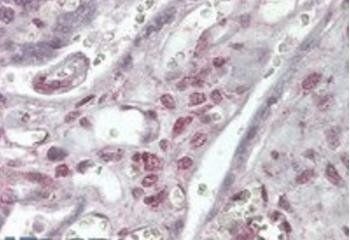
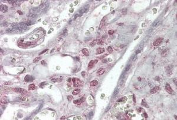
Immunohistochemical staining of Human Placenta using NHEJ1 antibody
3.8 µg/ml staining of paraffin embedded Human Placenta. Steamed antigen retrieval with citrate buffer pH6, AP-staining.
Primary incubation 1 hour at room temperature.Image A: HeLa nuclear cell lysate at primary Ab concentration 0.01 µg/ml, Images B, C, D: A431, Jurkat, K562 nuclear cell lysate at primary Ab concentration 0.03 µg/ml, Image E: HepG2 nuclear cell lysate at primary Ab concentration 0.1 µg/ml. (Loaded 35 µg protein in RIPA buffer, per lane). Detected by chemiluminescence.
Primary incubation 1 hour at room temperature. Image A: Human Skeletal muscle lysate at primary Ab concentration 0.03 ug/ml. (Loaded 35 µg protein in RIPA buffer, per lane). Detected by chemiluminescence.
Immunofluorescence analysis of paraformaldehyde fixed U2OS cells, permeabilized with 0.15% Triton. Primary incubation 1hr (10 ug/ml) followed by Alexa Fluor 488 secondary antibody (2 ug/ml), showing strong nuclear staining. Actin filaments were stained with phalloidin (red) and the nuclear stain is DAPI (blue). Negative control: Unimmunized goat IgG (10 ug/ml) followed by Alexa Fluor 488 secondary antibody (2 ug/ml).
Immunofluorescence analysis of paraformaldehyde fixed HepG2 cells, permeabilized with 0.15% Triton. Primary incubation 1hr (10 ug/ml) followed by Alexa Fluor 488 secondary antibody (2 ug/ml), showing nuclear and cytoplasmic staining. Actin filaments were stained with phalloidin (red) and the nuclear stain is DAPI (blue). Negative control: Unimmunized goat IgG (10 ug/ml) followed by Alexa Fluor 488 secondary antibody (2 ug/ml).
Flow cytometric analysis of paraformaldehyde fixed HepG2 cells (blue line), permeabilized with 0.5% Triton. Primary incubation 1hr (10 ug/ml) followed by Alexa Fluor 488 secondary antibody (1 ug/ml). IgG control: Unimmunized goat IgG (black line) followed by Alexa Fluor 488 secondary antibody.
Western blot analysis of Human Thyroid lysate using NHEJ1 antibody
* VAT and and shipping costs not included. Errors and price changes excepted