Purified polyclonal antibody supplied in PBS with 0.09% (W/V) sodium azide. This antibody is purified through a protein A column, followed by peptide affinity purification.
Application Dilute:
IHC-P - 1:100-500, WB - 1:2000
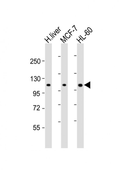
All lanes: Anti-RSBN1 Antibody (N-term) at 1:2000 dilution. Lane 1: human liver lysate. Lane 2: MCF-7 whole cell lysate. Lane 3: HL-60 whole cell lysate. Lysates/proteins at 20 µg per lane. Secondary Goat Anti-Rabbit IgG, (H+L), Peroxidase conjugated at 1/10000 dilution. Predicted band size: 90 kDa. Blocking/Dilution buffer: 5% NFDM/TBST.
RSBN1 Antibody (N-term) western blot analysis in Jurkat cell line lysates (35 ug/lane). This demonstrates the RSBN1 antibody detected the RSBN1 protein (arrow).
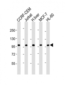
All lanes: Anti-RSBN1 Antibody (N-term) at 1:2000 dilution. Lane 1: CCRF-CEM whole cell lysate. Lane 2: Jurkat whole cell lysate. Lane 3: human liver lysate. Lane 4: MCF-7 whole cell lysate. Lane 5: HL-60 whole cell lysate.Lysates/proteins at 20 µg per lane. Secondary Goat Anti-Rabbit IgG, (H+L), Peroxidase conjugated at 1/10000 dilution. Predicted band size: 90 kDa. Blocking/Dilution buffer: 5% NFDM/TBST.
RSBN1 Antibody (N-term) immunohistochemistry analysis in formalin fixed and paraffin embedded human plecenta tissue followed by peroxidase conjugation of the secondary antibody and DAB staining. This data demonstrates the use of RSBN1 Antibody (N-term) for immunohistochemistry. Clinical relevance has not been evaluated.
Staining RSBN1 in human kidney tissue sections by Immunohistochemistry (IHC-P - paraformaldehyde-fixed, paraffin-embedded sections). Tissue was fixed with formaldehyde and blocked with 3% BSA for 0.5 hour at room temperature, antigen retrieval was by heat mediation with a citrate buffer (pH6). Samples were incubated with primary antibody (1/25) for 1 hours at 37C. A undiluted biotinylated goat polyvalent antibody was used as the secondary Antibody.
Staining RSBN1 in human colon tissue sections by Immunohistochemistry (IHC-P - paraformaldehyde-fixed, paraffin-embedded sections). Tissue was fixed with formaldehyde and blocked with 3% BSA for 0.5 hour at room temperature, antigen retrieval was by heat mediation with a citrate buffer (pH6). Samples were incubated with primary antibody (1/25) for 1 hours at 37C. A undiluted biotinylated goat polyvalent antibody was used as the secondary Antibody.
RSBN1 Antibody (N-term) flow cytometric analysis of Jurkat cells (right histogram) compared to a negative control cell (left histogram). FITC-conjugated donkey-anti-rabbit secondary antibodies were used for the analysis.
* VAT and and shipping costs not included. Errors and price changes excepted