Purified polyclonal antibody supplied in PBS with 0.09% (W/V) sodium azide. This antibody is purified through a protein A column, followed by peptide affinity purification.
Application Dilute:
WB - 1:1000, IHC-P - 1:100-500, FC - 1:10-50
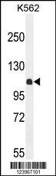
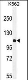
KIAA1324L Antibody (N-term) western blot analysis in K562 cell line lysates (35 ug/lane). This demonstrates the KIAA1324L antibody detected the KIAA1324L protein (arrow).
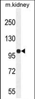
KIAA1324L Antibody (N-term) western blot analysis in mouse kidney tissue lysates (35 ug/lane). This demonstrates the KIAA1324L antibody detected the KIAA1324L protein (arrow).
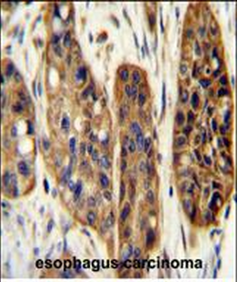
KIAA1324L Antibody (N-term) immunohistochemistry analysis in formalin fixed and paraffin embedded human esophagus carcinoma followed by peroxidase conjugation of the secondary antibody and DAB staining. This data demonstrates the use of the KIAA1324L Antibody (N-term) for immunohistochemistry. Clinical relevance has not been evaluated.
KIAA1324L Antibody (N-term) flow cytometric analysis of K562 cells (right histogram) compared to a negative control cell (left histogram). FITC-conjugated goat-anti-rabbit secondary antibodies were used for the analysis.
KIAA1324L Antibody (N-term) western blot analysis in K562 cell line lysates (35 ug/lane). This demonstrates the KIAA1324L antibody detected the KIAA1324L protein (arrow).
KIAA1324L Antibody (N-term) western blot analysis in mouse kidney tissue lysates (35 ug/lane). This demonstrates the KIAA1324L antibody detected the KIAA1324L protein (arrow).
KIAA1324L Antibody (N-term) immunohistochemistry analysis in formalin fixed and paraffin embedded human esophagus carcinoma followed by peroxidase conjugation of the secondary antibody and DAB staining. This data demonstrates the use of the KIAA1324L Antibody (N-term) for immunohistochemistry. Clinical relevance has not been evaluated.
KIAA1324L Antibody (N-term) flow cytometric analysis of K562 cells (right histogram) compared to a negative control cell (left histogram). FITC-conjugated goat-anti-rabbit secondary antibodies were used for the analysis.
* VAT and and shipping costs not included. Errors and price changes excepted