Purified monoclonal antibody supplied in PBS with 0.09% (W/V) sodium azide. This antibody is purified through a protein G column, followed by dialysis against PBS.
Application Dilute:
WB - 1:8000
All lanes: Anti-FASN Antibody (Center) at 1:500-1:2000 dilution. Lane 1: A549 whole cell lysate. Lane 2: Hela whole cell lysate. Lane 3: 293 whole cell lysate. Lane 4: Ramos whole cell lysate. Lane 5: HepG2 whole cell lysate. Lysates/proteins at 20 µg per lane. Secondary Goat Anti-mouse IgG, (H+L), Peroxidase conjugated at 1/10000 dilution. Predicted band size: 273 kDa. Blocking/Dilution buffer: 5% NFDM/TBST.
Fluorescent confocal image of HepG2 cells stained with FASN (Center) Antibody. HepG2 cells were fixed with 4% PFA (20 min), permeabilized with Triton X-100 (0.2%, 30 min). Cells were then incubated with FASN primary antibody (1:200, 2 h at room temperature). For secondary antibody, Alexa Fluor 488 conjugated donkey anti-mouse antibody (green) was used (1:1000, 1h). Nuclei were counterstained with Hoechst 33342 (blue) (10 µg/ml, 5 min).
Immunofluorescent analysis of 4% paraformaldehyde-fixed, 0. 1% Triton X-100 permeabilized U-2 OS ((human cervical epithelial adenocarcinoma cell line) cells labeling FASN at 1/25 dilution, followed by Dylight 488-conjugated goat anti-mouse IgG (35503) secondary antibody at 1/200 dilution (green). Immunofluorescence image showing cytoplasm Hela cell line. The nuclear counter stain is DAPI (blue).
Immunofluorescent analysis of 4% paraformaldehyde-fixed, 0. 1% Triton X-100 permeabilized HepG2 (human liver hepatocellular carcinoma cell line) cells labeling FASN at 1/25 dilution, followed by Dylight 488-conjugated goat anti-mouse IgG (35503) secondary antibody at 1/200 dilution (green). Immunofluorescence image showing cytoplasm HepG2 cell line. The nuclear counter stain is DAPI (blue).
FASN Antibody (Center) western blot analysis in mouse brain tissue lysates (35 µg/lane). This demonstrates the FASN (Center) antibody detected the FASN (Center) protein (arrow).
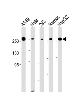
All lanes: Anti-FASN Antibody (Center) at 1:8000 dilution. Lane 1: A549 whole cell lysate. Lane 2: Hela whole cell lysate. Lane 3: 293 whole cell lysate. Lane 4: Ramos whole cell lysate. Lane 5: HepG2 whole cell lysate.Lysates/proteins at 20 µg per lane. Secondary Goat Anti-mouse IgG, (H+L), Peroxidase conjugated at 1/10000 dilution. Predicted band size: 273 kDa. Blocking/Dilution buffer: 5% NFDM/TBST.
Anti- at 1:1000 dilution + HepG2 whole cell lysate.Lysates/proteins at 20 µg per lane. Secondary Goat Anti-mouse IgG, (H+L), Peroxidase conjugated at 1/10000 dilution. Predicted band size: 273 kDa. Blocking/Dilution buffer: 5% NFDM/TBST.
* VAT and and shipping costs not included. Errors and price changes excepted