Purified monoclonal antibody supplied in PBS with 0.09% (W/V) sodium azide. This antibody is purified through a protein G column, followed by dialysis against PBS.
Application Dilute:
IF - 1:10-50, WB - 1:100-500, IHC-P - 1:100-500, FC - 1:10-50
CD44 antibody confocal immunofluorescent analysis with hela cell. 0.01 mg/ml primary antibody was followed by PE-conjugated goat anti-mouse lgG (whole molecule). PE emits red fluorescence. DAPI was used to stain the cell nuclear (blue).
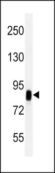
All lanes: Anti-CD44 Antibody at 1:2000 dilution. Lane 1: Hela whole cell lysate. Lane 2: HUVEC whole cell lysate.Lysates/proteins at 20 µg per lane. Secondary Goat Anti-mouse IgG, (H+L), Peroxidase conjugated at 1/10000 dilution. Predicted band size: 82 kDa. Blocking/Dilution buffer: 5% NFDM/TBST.
CD44 antibody immunohistochemistry analysis in formalin fixed and paraffin embedded human esophagus carcinoma followed by peroxidase conjugation of the secondary antibody and DAB staining. This data demonstrates the use of the CD44 antibody for immunohistochemistry. Clinical relevance has not been evaluated.
Staining CD44 in human skin tissue sections by Immunohistochemistry (IHC-P - paraformaldehyde-fixed, paraffin-embedded sections). Tissue was fixed with formaldehyde and blocked with 3% BSA for 0.5 hour at room temperature, antigen retrieval was by heat mediation with a citrate buffer (pH6). Samples were incubated with primary antibody (1/25) for 1 hours at 37C. A undiluted biotinylated goat polyvalent antibody was used as the secondary Antibody.
Staining CD44 in human lung adenocarcinoma tissue sections by Immunohistochemistry (IHC-P - paraformaldehyde-fixed, paraffin-embedded sections). Tissue was fixed with formaldehyde and blocked with 3% BSA for 0.5 hour at room temperature, antigen retrieval was by heat mediation with a citrate buffer (pH6). Samples were incubated with primary antibody (1/25) for 1 hours at 37C. A undiluted biotinylated goat polyvalent antibody was used as the secondary Antibody.
* VAT and and shipping costs not included. Errors and price changes excepted