Purified monoclonal antibody supplied in PBS with 0.09% (W/V) sodium azide. This antibody is purified through a protein G column, followed by dialysis against PBS.
Application Dilute:
WB - 1:1000
All lanes: Anti-ACTB Antibody at 1:2000 dilution + C6 cell lysate. Lysates/proteins at 20 µg per lane. Secondary Goat Anti-Mouse IgG, (H+L), Peroxidase conjugated at 1/10000 dilution. Predicted band size: 41 kDa. Blocking/Dilution buffer: 5% NFDM/TBST.
Confocal immunofluorescent analysis of ACTB Antibody with Hela cell followed by Alexa Fluor 488-conjugated goat anti-mouse lgG (green). DAPI was used to stain the cell nuclear (blue).
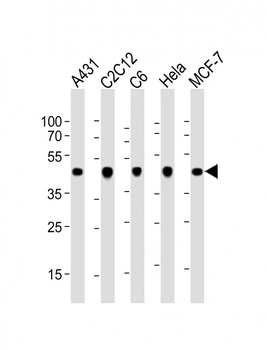
All lanes: Anti-ACTB Antibody at 1:1000 dilution. Lane 1: A431 whole cell lysate. Lane 2: C2C12 whole cell lysate. Lane 3: C6 whole cell lysate. Lane 4: Hela whole cell lysate. Lane 5: MCF-7 whole cell lysate.Lysates/proteins at 20 µg per lane. Secondary Goat Anti-mouse IgG, (H+L), Peroxidase conjugated at 1/10000 dilution. Predicted band size: 42 kDa. Blocking/Dilution buffer: 5% NFDM/TBST.
Western blot analysis of anti-ACTB Antibody in K562, HL-60, Hela cell line, mouse spleen, mouse liver tissue lysates, mouse NIH-3T3 cell line lysate and mouse cerebellum, mouse brain tissue lysates (35 µg/lane). ACTB (arrow) was detected using the purified Mab.
All lanes: Anti-ACTB Antibody at 1:1000 dilution. Lane 1: Hela whole cell lysate. Lane 2: HepG2 whole cell lysate. Lane 3: NIH-3T3 whole cell lysate.Lysates/proteins at 20 µg per lane. Secondary Goat Anti-mouse IgG, (H+L), Peroxidase conjugated at 1/10000 dilution. Predicted band size: 42 kDa. Blocking/Dilution buffer: 5% NFDM/TBST.
Staining ACTB in human heart tissue sections by Immunohistochemistry (IHC-P - paraformaldehyde-fixed, paraffin-embedded sections). Tissue was fixed with formaldehyde and blocked with 3% BSA for 0.5 hour at room temperature, antigen retrieval was by heat mediation with a citrate buffer (pH6). Samples were incubated with primary antibody (1/25) for 1 hours at 37C. A undiluted biotinylated goat polyvalent antibody was used as the secondary Antibody.
Immunohistochemical analysis of paraffin-embedded H.skeletal muscle section using Beta-Actin Antibody. diluted at 1:25 dilution. A peroxidase-conjugated goat anti-mouse IgG at 1:400 dilution was used as the secondary antibody, followed by DAB staining.
* VAT and and shipping costs not included. Errors and price changes excepted