E.coli-derived human AATF recombinant protein (Position: Q5-H552). Human AATF shares 78.5% and 79.9% amino acid (aa) sequence identity with mouse and rat AATF, respectively.
Conjugation:
Unconjugated
Alternative Names:
AATF, CHE1, DED, HSPC277, Protein AATF, Apoptosis-antagonizing transcription factor, Rb-binding protein Che-1
Anti-AATF Antibody. Tested in WB, IHC, ICC/IF, IF, ELISA applications. This antibody reacts with Human, Mouse, Rat.
Clonality:
Polyclonal
Concentration:
Adding 0.2 ml of distilled water will yield a concentration of 500 µg/ml.
Western blot, 0.25-0.5 µg/ml, Human Immunohistochemistry (Paraffin-embedded Section), 1-2 µg/ml, Human, Mouse Immunocytochemistry/Immunofluorescence, 5 µg/ml, Human Immunofluorescence, 5 µg/ml, Rat ELISA, 0.1-0.5 µg/ml, -
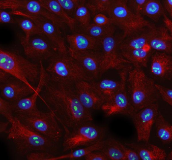
IF analysis of AATF using anti-AATF antibody and anti-Beta Tubulin antibody. AATF was detected in immunocytochemical section of A549 cell. Enzyme antigen retrieval was performed using IHC enzyme antigen retrieval reagent for 15 mins. The cells were blocked with 10% goat serum. And then incubated with 5 µg/mL rabbit anti-AATF Antibody and mouse anti-Beta Tubulin antibody overnight at 4C. DyLight488 Conjugated Goat Anti-Rabbit IgG and Cy3 Conjugated Goat Anti-Mouse IgG were used as secondary antibody at 1:500 dilution and incubated for 30 minutes at 37C. The section was counterstained with DAPI. Visualize using a fluorescence microscope and filter sets appropriate for the label used.
IF analysis of AATF using anti-AATF antibody. AATF was detected in a paraffin-embedded section of rat brain tissue. Heat mediated antigen retrieval was performed in EDTA buffer (pH8.0, epitope retrieval solution). The tissue section was blocked with 10% goat serum. The tissue section was then incubated with 5 µg/mL rabbit anti-AATF Antibody overnight at 4C. DyLight594 Conjugated Goat Anti-Rabbit IgG was used as secondary antibody at 1:500 dilution and incubated for 30 minutes at 37C. The section was counterstained with DAPI. Visualize using a fluorescence microscope and filter sets appropriate for the label used.
IHC analysis of AATF using anti-AATF antibody. AATF was detected in a paraffin-embedded section of mouse cerebellum tissue. Heat mediated antigen retrieval was performed in EDTA buffer (pH8.0, epitope retrieval solution). The tissue section was blocked with 10% goat serum. The tissue section was then incubated with 2 µg/ml rabbit anti-AATF Antibody overnight at 4C. Peroxidase Conjugated Goat Anti-rabbit IgG was used as secondary antibody and incubated for 30 minutes at 37C. The tissue section was developed using HRP Conjugated Rabbit IgG Super Vision Assay Kit with DAB as the chromogen.
IHC analysis of AATF using anti-AATF antibody. AATF was detected in a paraffin-embedded section of mouse cerebellum tissue. Heat mediated antigen retrieval was performed in EDTA buffer (pH8.0, epitope retrieval solution). The tissue section was blocked with 10% goat serum. The tissue section was then incubated with 2 µg/ml rabbit anti-AATF Antibody overnight at 4C. Peroxidase Conjugated Goat Anti-rabbit IgG was used as secondary antibody and incubated for 30 minutes at 37C. The tissue section was developed using HRP Conjugated Rabbit IgG Super Vision Assay Kit with DAB as the chromogen.
IHC analysis of AATF using anti-AATF antibody. AATF was detected in a paraffin-embedded section of mouse cerebellum tissue. Heat mediated antigen retrieval was performed in EDTA buffer (pH8.0, epitope retrieval solution). The tissue section was blocked with 10% goat serum. The tissue section was then incubated with 2 µg/ml rabbit anti-AATF Antibody overnight at 4C. Peroxidase Conjugated Goat Anti-rabbit IgG was used as secondary antibody and incubated for 30 minutes at 37C. The tissue section was developed using HRP Conjugated Rabbit IgG Super Vision Assay Kit with DAB as the chromogen.
IHC analysis of AATF using anti-AATF antibody. AATF was detected in a paraffin-embedded section of mouse cerebellum tissue. Heat mediated antigen retrieval was performed in EDTA buffer (pH8.0, epitope retrieval solution). The tissue section was blocked with 10% goat serum. The tissue section was then incubated with 2 µg/ml rabbit anti-AATF Antibody overnight at 4C. Peroxidase Conjugated Goat Anti-rabbit IgG was used as secondary antibody and incubated for 30 minutes at 37C. The tissue section was developed using HRP Conjugated Rabbit IgG Super Vision Assay Kit with DAB as the chromogen.
Western blot analysis of AATF using anti-AATF antibody. Electrophoresis was performed on a 5-20% SDS-PAGE gel at 70V (Stacking gel) / 90V (Resolving gel) for 2-3 hours. The sample well of each lane was loaded with 30 ug of sample under reducing conditions. Lane 1: human 293T whole cell lysates, L
* VAT and and shipping costs not included. Errors and price changes excepted