E.coli-derived human HDHD2 recombinant protein (Position: M1-L259). Human HDHD2 shares 89.2% and 90% amino acid (aa) sequence identity with mouse and rat HDHD2, respectively.
Conjugation:
Unconjugated
Alternative Names:
3110052N05Rik, HDHD2
Anti-HDHD2 Antibody. Tested in ELISA, IHC, WB, Flow Cytometry applications. This antibody reacts with Human, Mouse, Rat.
Clonality:
Polyclonal
Concentration:
Adding 0.2 ml of distilled water will yield a concentration of 500 µg/ml.
Haloacid dehalogenase-like hydrolase domain-containing protein 2
Application Dilute:
Western blot, 0.25-0.5 µg/ml, Human, Mouse, Rat Immunohistochemistry (Paraffin-embedded Section), 2-5 µg/ml, Human Flow Cytometry (Fixed), 1-3 µg/1x10 6 cells, Human ELISA, 0.1-0.5 µg/ml, -
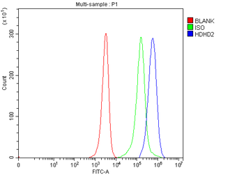
Flow Cytometry analysis of JK cells using anti-HDHD2 antibody. Overlay histogram showing JK cells (Blue line). The cells were fixed with 4% paraformaldehyde and blocked with 10% normal goat serum. And then incubated with rabbit anti-HDHD2 Antibody (1 µg/1x10 6 cells) for 30 min at 20C. DyLight488 conjugated goat anti-rabbit IgG (5-10 µg/1x10 6 cells) was used as secondary antibody for 30 minutes at 20C. Isotype control antibody (Green line) was rabbit IgG (1 µg/1x10 6) used under the same conditions. Unlabelled sample (Red line) was also used as a control.
IHC analysis of HDHD2 using anti-HDHD2 antibody. HDHD2 was detected in a paraffin-embedded section of human colon adenocarcinoma tissue. Heat mediated antigen retrieval was performed in EDTA buffer (pH8.0, epitope retrieval solution). The tissue section was blocked with 10% goat serum. The tissue section was then incubated with 2 µg/ml rabbit anti-HDHD2 Antibody overnight at 4C. Peroxidase Conjugated Goat Anti-rabbit IgG was used as secondary antibody and incubated for 30 minutes at 37C. The tissue section was developed using HRP Conjugated Rabbit IgG Super Vision Assay Kit with DAB as the chromogen.
IHC analysis of HDHD2 using anti-HDHD2 antibody. HDHD2 was detected in a paraffin-embedded section of human colon adenocarcinoma tissue. Heat mediated antigen retrieval was performed in EDTA buffer (pH8.0, epitope retrieval solution). The tissue section was blocked with 10% goat serum. The tissue section was then incubated with 2 µg/ml rabbit anti-HDHD2 Antibody overnight at 4C. Peroxidase Conjugated Goat Anti-rabbit IgG was used as secondary antibody and incubated for 30 minutes at 37C. The tissue section was developed using HRP Conjugated Rabbit IgG Super Vision Assay Kit with DAB as the chromogen.
IHC analysis of HDHD2 using anti-HDHD2 antibody. HDHD2 was detected in a paraffin-embedded section of human pancreas ductal adenocarcinoma tissue. Heat mediated antigen retrieval was performed in EDTA buffer (pH8.0, epitope retrieval solution). The tissue section was blocked with 10% goat serum. The tissue section was then incubated with 2 µg/ml rabbit anti-HDHD2 Antibody overnight at 4C. Peroxidase Conjugated Goat Anti-rabbit IgG was used as secondary antibody and incubated for 30 minutes at 37C. The tissue section was developed using HRP Conjugated Rabbit IgG Super Vision Assay Kit with DAB as the chromogen.
IHC analysis of HDHD2 using anti-HDHD2 antibody. HDHD2 was detected in a paraffin-embedded section of human pancreas ductal adenocarcinoma tissue. Heat mediated antigen retrieval was performed in EDTA buffer (pH8.0, epitope retrieval solution). The tissue section was blocked with 10% goat serum. The tissue section was then incubated with 2 µg/ml rabbit anti-HDHD2 Antibody overnight at 4C. Peroxidase Conjugated Goat Anti-rabbit IgG was used as secondary antibody and incubated for 30 minutes at 37C. The tissue section was developed using HRP Conjugated Rabbit IgG Super Vision Assay Kit with DAB as the chromogen.
IHC analysis of HDHD2 using anti-HDHD2 antibody. HDHD2 was detected in a paraffin-embedded section of human testicular seminoma tissue. Heat mediated antigen retrieval was performed in EDTA buffer (pH8.0, epitope retrieval solution). The tissue section was blocked with 10% goat serum. The tissue section was then incubated with 2 µg/ml rabbit anti-HDHD2 Antibody overnight at 4C. Peroxidase Conjugated Goat Anti-rabbit IgG was used as secondary antibody and incubated for 30 minutes at 37C. The tissue section was developed using HRP Conjugated Rabbit IgG Super Vision Assay Kit with DAB as the chromogen.
IHC analysis of HDHD2 using anti-HDHD2 antibody. HDHD2 was detected in a paraffin-embedded section of human testicular seminoma tissue. Heat mediated antigen retrieval was performed in EDTA buffer (pH8.0, epitope retrieval solution). The tissue section was blocked with 10% goat serum. The tissue section was then incubated with 2 µg/ml rabbit anti-HDHD2 Antibody
* VAT and and shipping costs not included. Errors and price changes excepted