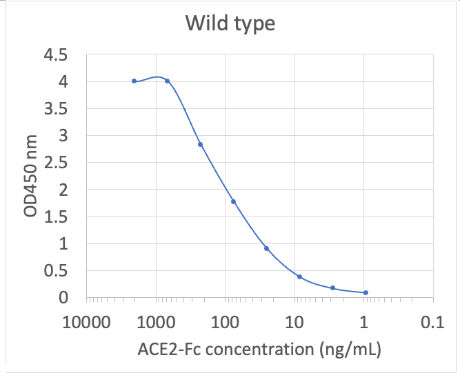
Microtiter wells were coated with 100 uL of each spike trimer at 2 ug/mL in PBS at 4?C overnight. The wells were washed with PBS and blocked with 200 µL of 1% BSA/PBS. ACE2-Fc was serially diluted from 2 µg/mL in 1% BSA/PBS. The blocker was discarded, and the wells were incubated with 100 µL of serially diluted ACE2-Fc at 37?C for 1 hour. The wells were washed with PBS and the bound ACE2-Fc was detected with 100 µL of Peroxidase AffiniPure Goat Anti-Human IgG, Fc? fragment specific (1:5, 000 in 1% BSA/PBS) at 37?C for 1 hour. The wells were washed with PBS and the wells were developed with 100 µL of MB/E Ultra Sensitive, Blue, Horseradish Peroxidase Substrate at RT for 5 min. The reaction was stopped with 100 µL of 0.6N H2SO4 and the signals were read at 450 nm using a plate reader.
Microtiter wells were coated with 100 uL of each spike trimer at 2 ug/mL in PBS at 4?C overnight. The wells were washed with PBS and blocked with 200 µL of 1% BSA/PBS. ACE2-Fc was serially diluted from 2 µg/mL in 1% BSA/PBS. The blocker was discarded, and the wells were incubated with 100 µL of serially diluted ACE2-Fc at 37?C for 1 hour. The wells were washed with PBS and the bound ACE2-Fc was detected with 100 µL of Peroxidase AffiniPure Goat Anti-Human IgG, Fcgamma fragment specific (1:5, 000 in 1% BSA/PBS) at 37?C for 1 hour. The wells were washed with PBS and the wells were developed with 100 µL of MB/E Ultra Sensitive, Blue, Horseradish Peroxidase Substrate at RT for 5 min. The reaction was stopped with 100 µL of 0.6N H2SO4 and the signals were read at 450 nm using a plate reader.
Microtiter wells were coated with 100 uL of each spike trimer at 2 ug/mL in PBS at 4?C overnight. The wells were washed with PBS and blocked with 200 µL of 1% BSA/PBS. ACE2-Fc was serially diluted from 2 µg/mL in 1% BSA/PBS. The blocker was discarded, and the wells were incubated with 100 µL of serially diluted ACE2-Fc at 37?C for 1 hour. The wells were washed with PBS and the bound ACE2-Fc was detected with 100 µL of Peroxidase AffiniPure Goat Anti-Human IgG, Fc? fragment specific (1:5, 000 in 1% BSA/PBS) at 37?C for 1 hour. The wells were washed with PBS and the wells were developed with 100 µL of MB/E Ultra Sensitive, Blue, Horseradish Peroxidase Substrate at RT for 5 min. The reaction was stopped with 100 µL of 0.6N H2SO4 and the signals were read at 450 nm using a plate reader.
Microtiter wells were coated with 100 uL of each spike trimer at 2 ug/mL in PBS at 4?C overnight. The wells were washed with PBS and blocked with 200 µL of 1% BSA/PBS. ACE2-Fc was serially diluted from 2 µg/mL in 1% BSA/PBS. The blocker was discarded, and the wells were incubated with 100 µL of serially diluted ACE2-Fc at 37?C for 1 hour. The wells were washed with PBS and the bound ACE2-Fc was detected with 100 µL of Peroxidase AffiniPure Goat Anti-Human IgG, Fc? fragment specific (1:5, 000 in 1% BSA/PBS) at 37?C for 1 hour. The wells were washed with PBS and the wells were developed with 100 µL of MB/E Ultra Sensitive, Blue, Horseradish Peroxidase Substrate at RT for 5 min. The reaction was stopped with 100 µL of 0.6N H2SO4 and the signals were read at 450 nm using a plate reader.
Microtiter wells were coated with 100 uL of each spike trimer at 2 ug/mL in PBS at 4?C overnight. The wells were washed with PBS and blocked with 200 µL of 1% BSA/PBS. ACE2-Fc was serially diluted from 2 µg/mL in 1% BSA/PBS. The blocker was discarded, and the wells were incubated with 100 µL of serially diluted ACE2-Fc at 37?C for 1 hour. The wells were washed with PBS and the bound ACE2-Fc was detected with 100 µL of Peroxidase AffiniPure Goat Anti-Human IgG, Fc? fragment specific (1:5, 000 in 1% BSA/PBS) at 37?C for 1 hour. The wells were washed with PBS and the wells were developed with 100 µL of MB/E Ultra Sensitive, Blue, Horseradish Peroxidase Substrate at RT for 5 min. The reaction was stopped with 100 µL of 0.6N H2SO4 and the signals were read at 450 nm using a plate reader.
Microtiter wells were coated
* VAT and and shipping costs not included. Errors and price changes excepted