
beta Amyloid/APP Rabbit Polyclonal Antibody, Unconjugated
Catalog Number:
BYT-ORB196261
- Images (8)


| Article Name: | beta Amyloid/APP Rabbit Polyclonal Antibody, Unconjugated |
| Biozol Catalog Number: | BYT-ORB196261 |
| Supplier Catalog Number: | orb196261 |
| Alternative Catalog Number: | BYT-ORB196261-100 |
| Manufacturer: | Biorbyt |
| Host: | Rabbit |
| Category: | Antikörper |
| Application: | ICC, IF, IHC, WB |
| Species Reactivity: | Human, Mouse, Rat |
| Immunogen: | A synthetic peptide corresponding to a sequence at the C-terminus of human APP, different from the related mouse and rat sequences by three amino acids. |
| Conjugation: | Unconjugated |
| Alternative Names: | Amyloid beta A4 protein, ABPP, APPI, APP, Alzheimer disease amyloid protein, Cerebral vascular amyloid peptide, CVAP, PreA4, Protease nexin-II, PN-II, N-APP, Soluble APP-alpha, S-APP-alpha, Soluble APP-beta, S-APP-beta, C99, Beta-amyloid protein 42, Beta-APP42, Beta-amyloid protein 40, Beta-APP40, C83, P3 (42), P3 (40), C80, Gamma-secretase C-terminal fragment 59, Amyloid intracellular domain 59, AICD-59, AID (59), Gamma-CTF (59), Gamma-secretase C-terminal fragment 57, Amyloid intracellular domain 57, AICD-57, AID (57), Gamma-CTF (57), Gamma-secretase C-terminal fragment 50, Amyloid intracellular domain 50, AICD-50, AID (50), Gamma-CTF (50), C31, APP, A4, AD1 |
| beta Amyloid/APP Rabbit Polyclonal Antibody |
| Clonality: | Polyclonal |
| Concentration: | Adding 0.2 ml of distilled water will yield a concentration of 500 µg/ml. |
| Molecular Weight: | 120 kDa |
| UniProt: | P05067 |
| Buffer: | Each vial contains antibody formulated with stabilizing components, 0.9mg NaCl, 0.2mg Na2HPO4, 0.01mg NaN3. *This antibody is supplied in a stabilized formulation. Compatibility with conjugation reactions depends on the chemistry of the conjugation method |
| Form: | Lyophilized |
| Target: | Amyloid-beta precursor protein |
| Application Dilute: | Western blot, 0.1-0.5µg/ml, Human, Mouse, Rat Immunohistochemistry (Paraffin-embedded Section), 0.5-1µg/ml, Mouse, Rat, Human Immunocytochemistry/Immunofluorescence, 2µg/ml, Human |
|
|
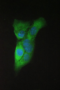
IF analysis of APP using anti-APP antibody. APP was detected in immunocytochemical section of A431 cell. Enzyme antigen retrieval was performed using IHC enzyme antigen retrieval reagent for 15 mins. The cells were blocked with 10% goat serum. And then incubated with 2 µg/mL rabbit anti-APP Antibody overnight at 4C. DyLight488 Conjugated Goat Anti-Rabbit IgG was used as secondary antibody at 1:100 dilution and incubated for 30 minutes at 37C. The section was counterstained with DAPI. Visualize using a fluorescence microscope and filter sets appropriate for the label used. |
|
|
IF analysis of APP using anti-APP antibody.APP was detected in immunocyt |
|
|
IHC analysis of APP using anti-APP antibody. APP was detected in paraffin-embedded section of human glioma tissue. Heat mediated antigen retrieval was performed in citrate buffer (pH6, epitope retrieval solution) for 20 mins. The tissue section was blocked with 10% goat serum. The tissue section was then incubated with 1 µg/ml rabbit anti-APP Antibody overnight at 4C. Biotinylated goat anti-rabbit IgG was used as secondary antibody and incubated for 30 minutes at 37C. The tissue section was developed using Strepavidin-Biotin-Complex (SABC) with DAB as the chromogen. |
|
|
IHC analysis of APP using anti-APP antibody. APP was detected in paraffin-embedded section of human renal cancer tissue. Heat mediated antigen retrieval was performed in citrate buffer (pH6, epitope retrieval solution) for 20 mins. The tissue section was blocked with 10% goat serum. The tissue section was then incubated with 1 µg/ml rabbit anti-APP Antibody overnight at 4C. Biotinylated goat anti-rabbit IgG was used as secondary antibody and incubated for 30 minutes at 37C. The tissue section was developed using Strepavidin-Biotin-Complex (SABC) with DAB as the chromogen. |
|
|
IHC analysis of APP using anti-APP antibody. APP was detected in paraffin-embedded section of human tonsil tissue. Heat mediated antigen retrieval was performed in citrate buffer (pH6, epitope retrieval solution) for 20 mins. The tissue section was blocked with 10% goat serum. The tissue section was then incubated with 1 µg/ml rabbit anti-APP Antibody overnight at 4C. Biotinylated goat anti-rabbit IgG was used as secondary antibody and incubated for 30 minutes at 37C. The tissue section was developed using Strepavidin-Biotin-Complex (SABC) with DAB as the chromogen. |
|
|
IHC analysis of APP using anti-APP antibody. APP was detected in paraffin-embedded section of mouse brain tissue. Heat mediated antigen retrieval was performed in citrate buffer (pH6, epitope retrieval solution) for 20 mins. The tissue section was blocked with 10% goat serum. The tissue section was then incubated with 1 µg/ml rabbit anti-APP Antibody overnight at 4C. Biotinylated goat anti-rabbit IgG was used as secondary antibody and incubated for 30 minutes at 37C. The tissue section was developed using Strepavidin-Biotin-Complex (SABC) with DAB as the chromogen. |
|
|
IHC analysis of APP using anti-APP antibody. APP was detected in paraffin-embedded section of rat brain tissue. Heat mediated antigen retrieval was performed in citrate buffer (pH6, epitope retrieval solution) for 20 mins. The tissue section was blocked with 10% goat serum. The tissue section was then incubated with 1 µg/ml rabbit anti-APP Antibody overnight at 4C. Biotinylated goat anti-rabbit IgG was used as secondary antibody and incubated for 30 minutes at 37C. The tissue section was developed using Strepavidin-Biotin-Complex (SABC) with DAB as the chromogen. |
|
|
Western blot analysis of APP using anti-APP antibody. Electrophoresis was performed on a 5-20% SDS-PAGE gel at 70V (Stacking gel) / 90V (Resolving gel) for 2-3 hours. The sample well of each lane was loaded with 50 ug of sample under reducing conditions. Lane 1: human Hela whole cell lysates, Lane 2: human U-87MG whole cell lysates, Lane 3: human T-47D whole cell lysates, Lane 4: human A549 whole cell lysates, Lane 5: human U2OS whole cell lysates, Lane 6: rat brain tissue lysates, Lane 7: mouse brain tissue lysates. After Electrophor |
Product Guarantee and Expert Support
