Peptide with sequence C-DEVNAFKQRKTA, from the internal region of the protein sequence according to NP_665798.1, NP_055944.2, NP_665801.1.
Conjugation:
Unconjugated
Alternative Names:
anti SEPT6 antibody, anti septin 6 antibody, anti KIAA0128 antibody, anti MGC16619 antibody, anti MGC20339 antibody, anti SEP2 antibody, anti SEPT2 antibody, anti septin 2 antibody
This product is a goat polyclonal antibody targeting SEPT6, supplied unconjugated at 0.5 mg/ml. Validated for multiple applications including ELISA, flow cytometry, immunofluorescence, immunohistochemistry, and Western blot, it exhibits reactivity with human samples and shows predicted reactivity with bovine and canine species.
Clonality:
Polyclonal
Molecular Weight:
48.9 kDa, 49.7 kDa, 49.2 kDa
Buffer:
Supplied at 0.5 mg/ml in Tris saline, 0.02% sodium azide, pH 7.3 with 0.5% bovine serum albumin. Aliquot and store at -20C. Minimize freezing and thawing.
Sequence:
DEVNAFKQRKTA
Target:
SEPT6
Application Dilute:
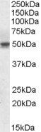
Peptide ELISA: antibody detection limit dilution 1:4000. Western blot: Approx 50kDa band observed in Human Testis, Duodenum, Kidney and Tonsil lysates, and in lysates of cell lines Daudi, Jurkat and MOLT-4 (calculated MW of 49.7kDa according to NP_055944.
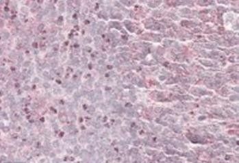
Immunohistochemical staining of Human Tonsil using 41888 antibody
Flow cytometric analysis of paraformaldehyde fixed A431 cells (blue line), permeabilized with 0.5% Triton. Primary incubation 1hr (10 ug/ml) followed by Alexa Fluor 488 secondary antibody (1 ug/ml). IgG control: Unimmunized goat IgG (black line) followed by Alexa Fluor 488 secondary antibody.
Immunofluorescence analysis of paraformaldehyde fixed A431 cells, permeabilized with 0.15% Triton. Primary incubation 1hr (10 ug/ml) followed by Alexa Fluor 488 secondary antibody (2 ug/ml), showing cytoplasmic staining. The nuclear stain is DAPI (blue). Negative control: Unimmunized goat IgG (10 ug/ml) followed by Alexa Fluor 488 secondary antibody (2 ug/ml).
Primary incubation 1 hour at room temperature. Image A: Human Testes lysate at primary Ab concentration 1 ug/ml, Image B: Human Duodenum lysate at primary Ab concentration 0.1 ug/ml, Image C: Human Kidney lysate at primary Ab concentration 0.5 ug/ml, Image D: Human Tonsil lysate at primary Ab concentration 0.3 ug/ml (Loaded 35 µg protein in RIPA buffer, per lane). Detected by chemiluminescence.
Primary incubation 1 hour at room temperature. Images A, B, C: Daudi, Jurkat, MOLT-4 cell lysates at primary Ab concentration 0.1 ug/ml (Loaded 35 µg protein in RIPA buffer, per lane). Detected by chemiluminescence.
3.8 µg/ml staining of paraffin embedded Human Tonsil. Steamed antigen retrieval with citrate buffer pH6, AP-staining.
Immunofluorescence analysis of paraformaldehyde fixed U2OS cells, permeabilized with 0.15% Triton. Primary incubation 1hr (10 ug/ml) followed by Alexa Fluor 488 secondary antibody (2 ug/ml), showing cytoplasmic staining. The nuclear stain is DAPI (blue). Negative control: Unimmunized goat IgG (10 ug/ml) followed by Alexa Fluor 488 secondary antibody (2 ug/ml).
Western blot analysis of Human Testis lysate using 41888 antibody
* VAT and and shipping costs not included. Errors and price changes excepted