Each vial contains antibody formulated with stabilizing components, 0.9 mg NaCl, 0.2 mg Na2HPO4, and 0.05 mg NaN3. *This antibody is supplied in a stabilized formulation. Compatibility with conjugation reactions depends on the chemistry of the conjugation
Form:
Lyophilized
Target:
von Willebrand factor
Application Dilute:
Western blot, 0.1-0.5µg/ml, Mouse, Rat Immunohistochemistry (Paraffin-embedded Section), 0.5-1µg/ml, Mouse, Rat Immunofluorescence, 2µg/ml, Rat
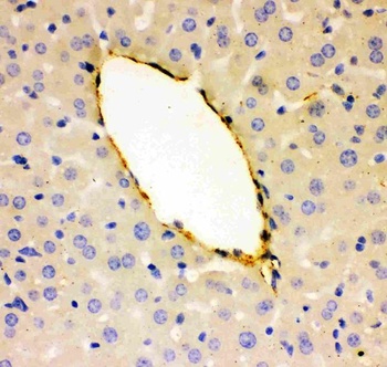
IHC(P) analysis of Mouse Liver Tissue using Anti-VWF Picoband antibody.
WB analysis of Recombinant Mouse VWF Protein 0.5ng using Anti-VWF Picoband antibody.
Anti-VWF Picoband antibody, Western blotting. All lanes: Anti VWF at 0.5 ug/ml. WB: Mouse Lung Tissue Lysate at 50 ug. Predicted bind size: 309 KD. Observed bind size: 309 KD.
Anti-VWF Picoband antibody, Western blotting. All lanes: Anti VWF at 0.5 ug/ml. WB: Recombinant Mouse VWF Protein 0.5 ng. Predicted bind size: 37 KD. Observed bind size: 37 KD.
IF analysis of VWF and alpha-Smooth Muscle Actin using anti-VWF antibody and anti-alpha-Smooth Muscle Actin antibody. VWF and alpha-Smooth Muscle Actin a paraffin-embedded section of rat lung tissue. Heat mediated antigen retrieval was performed in EDTA buffer (pH8.0, epitope retrieval solution). The tissue section was blocked with 10% goat serum. The tissue section was then incubated with 2 µg/mL rabbit anti-VWF antibody and mouse anti-alpha-Smooth Muscle Actin Antibody overnight at 4C. DyLight488 Conjugated Goat Anti-Rabbit IgG and Cy3 Conjugated Goat Anti-Mouse IgG were used as secondary antibody at 1:100 dilution and incubated for 30 minutes at 37C. The section was counterstained with DAPI. Visualize using a fluorescence microscope and filter sets appropriate for the label used.
IF analysis of VWF using anti-VWF antibody. VWF was detected in paraffin-embedded section of rat liver tissue. Heat mediated antigen retrieval was performed in EDTA buffer (pH8.0, epitope retrieval solution). The tissue section was blocked with 10% goat serum. The tissue section was then incubated with 2 µg/mL rabbit anti-VWF Antibody overnight at 4C. DyLight488 Conjugated Goat Anti-Rabbit IgG was used as secondary antibody at 1:100 dilution and incubated for 30 minutes at 37C. The section was counterstained with DAPI. Visualize using a fluorescence microscope and filter sets appropriate for the label used.
IHC analysis of VWF using anti-VWF antibody. VWF was detected in paraffin-embedded section of rat lung tissues. Heat mediated antigen retrieval was performed in citrate buffer (pH6, epitope retrieval solution) for 20 mins. The tissue section was blocked with 10% goat serum. The tissue section was then incubated with 1 µg/ml rabbit anti-VWF Antibody overnight at 4C. Biotinylated goat anti-rabbit IgG was used as secondary antibody and incubated for 30 minutes at 37C. The tissue section was developed using Strepavidin-Biotin-Complex (SABC) with DAB as the chromogen.
* VAT and and shipping costs not included. Errors and price changes excepted