E.coli-derived human Hsp60 recombinant protein (Position: A260-Q496). Human Hsp60 shares 97% amino acid (aa) sequence identity with both mouse and rat Hsp60.
Each vial contains 4 mg Trehalose, 0.9 mg NaCl and 0.2 mg Na2HPO4.
Form:
Lyophilized
Target:
60 kDa heat shock protein, mitochondrial
Application Dilute:
Western blot, 0.1-0.5µg/ml, Human, Mouse, Rat Immunohistochemistry (Paraffin-embedded Section), 0.5-1µg/ml, Human, Mouse, Rat Immunocytochemistry/Immunofluorescence, 2µg/ml, Human
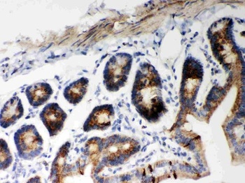
IHC(P) analysis of Mouse Intestine Tissue using Anti-Hsp60 Picoband antibody.
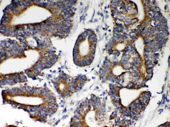
Anti-Hsp60 Picoband antibody, IHC(P): Human Intestinal Cancer Tissue.
IHC(P) analysis of Rat Intestine Tissue using Anti-Hsp60 Picoband antibody.
Anti-Hsp60 Picoband antibody, IHC(P): Rat Intestine Tissue.
IF analysis of Hsp60 using anti-Hsp60 antibody. Hsp60 was detected in immunocytochemical section of U20S cell. Enzyme antigen retrieval was performed using IHC enzyme antigen retrieval reagent for 15 mins. The cells were blocked with 10% goat serum. And then incubated with 2 µg/mL rabbit anti-Hsp60 Antibody overnight at 4C. DyLight488 Conjugated Goat Anti-Rabbit IgG was used as secondary antibody at 1:100 dilution and incubated for 30 minutes at 37C. The section was counterstained with DAPI. Visualize using a fluorescence microscope and filter sets appropriate for the label used.
IHC analysis of Hsp60 using anti-Hsp60 antibody. Hsp60 was detected in immunocytochemical section of SMMC-7721 Cell. Enzyme antigen retrieval was performed using IHC enzyme antigen retrieval reagent for 15 mins. The cells were blocked with 10% goat serum. And then incubated with 1 µg/ml rabbit anti-Hsp60 Antibody overnight at 4C. Biotinylated goat anti-rabbit IgG was used as secondary antibody and incubated for 30 minutes at 37C. The section was developed using Strepavidin-Biotin-Complex (SABC) with DAB as the chromogen.
Western blot analysis of Hsp60 using anti-Hsp60 antibody. Electrophoresis was performed on a 5-20% SDS-PAGE gel at 70V (Stacking gel) / 90V (Resolving gel) for 2-3 hours. The sample well of each lane was loaded with 30 ug of sample under reducing conditions. Lane 1: human Hela whole cell lysates, Lane 2: human A549 whole cell lysates, Lane 3: human PANC-1 whole cell lysates, Lane 4: human MCF-7 whole cell lysates, Lane 5: rat liver tissue lysates, Lane 6: mouse liver tissue lysates, Lane 7: mouse thymus tissue lysates. After electrophoresis, proteins were transferred to a nitrocellulose membrane at 150 mA for 50-90 minutes. Blocked the membrane with 5% non-fat milk/TBS for 1.5 hour at RT. The membrane was incubated with rabbit anti-Hsp60 antigen affinity purified polyclonal antibody at 0.5 µg/mL overnight at 4C, then washed with TBS-0.1% Tween 3 times with 5 minutes each and probed with a goat anti-rabbit IgG-HRP secondary antibody at a dilution of 1:5000 for 1.5 hour at RT. The signal is developed using an Enhanced Chemiluminescent detection (ECL) kit with Tanon 5200 system. A specific band was detected for Hsp60 at approximately 61 kDa. The expected band size for Hsp60 is at 61 kDa.
* VAT and and shipping costs not included. Errors and price changes excepted