Each vial contains antibody formulated with stabilizing components, 0.9 mg NaCl, 0.2 mg Na2HPO4, and 0.05 mg NaN3. *This antibody is supplied in a stabilized formulation. Compatibility with conjugation reactions depends on the chemistry of the conjugation
Form:
Lyophilized
Target:
Survival motor neuron protein
Application Dilute:
Western blot, 0.1-0.5µg/ml, Human, Mouse, Rat Immunohistochemistry (Paraffin-embedded Section), 0.5-1µg/ml, Human, Mouse, Rat Immunocytochemistry/Immunofluorescence, 2µg/ml, Human Flow Cytometry (Fixed), 1-3µg/1x10 6 cells, Human
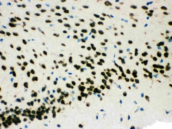
IHC(P) analysis of Mouse Brain Tissue using Anti-SMN1/2 Picoband antibody.
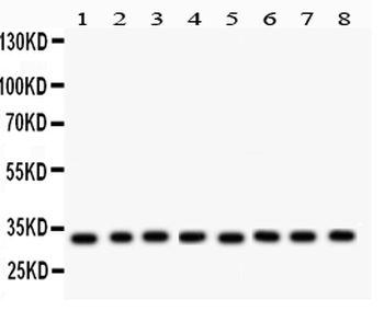
Anti-SMN1/2 Picoband antibody, Western blotting. All lanes: Anti SMN1/2 at 0.5 ug/ml. Lane 1: Rat Brain Tissue Lysate at 50 ug. Lane 2: Mouse Brain Tissue Lysate at 50 ug. Lane 3: Rat Liver Tissue Lysate at 50 ug. Lane 4: Mouse Liver Tissue Lysate at 50 ug. Lane 5: 293T Whole Cell Lysate at 40 ug. Lane 6: SMMC Whole Cell Lysate at 40 ug. Lane 7: HEPG2 Whole Cell Lysate at 40 ug. Lane 8: HELA Whole Cell Lysate at 40 ug. Predicted bind size: 32 KD. Observed bind size: 32 KD.
IHC(P) analysis of Rat Brain Tissue using Anti-SMN1/2 Picoband antibody.
Anti-SMN1/2 Picoband antibody, IHC(P): Human Mammary Cancer Tissue.
Anti-SMN1/2 Picoband antibody, IHC(P): Rat Brain Tissue.
Flow Cytometry analysis of A431 cells using anti-SMN1/2 antibody. Overlay histogram showing A431 cells (Blue line). To facilitate intracellular staining, cells were fixed with 4% paraformaldehyde and permeabilized with permeabilization buffer. The cells were blocked with 10% normal goat serum. And then incubated with rabbit anti-SMN1/2 Antibody (1 µg/1x10 6 cells) for 30 min at 20C. DyLight488 conjugated goat anti-rabbit IgG (5-10 µg/1x10 6 cells) was used as secondary antibody for 30 minutes at 20C. Isotype control antibody (Green line) was rabbit IgG (1 µg/1x10 6) used under the same conditions. Unlabelled sample without incubation with primary antibody and secondary antibody (Red line) was used as a blank control.
IF analysis of SMN1/2 using anti-SMN1/2 antibody. SMN1/2 was detected in immunocytochemical section of U20S cells. Enzyme antigen retrieval was performed using IHC enzyme antigen retrieval reagent for 15 mins. The cells were blocked with 10% goat serum. And then incubated with 2 µg/mL rabbit anti-SMN1/2 Antibody overnight at 4C. DyLight488 Conjugated Goat Anti-Rabbit IgG was used as secondary antibody at 1:100 dilution and incubated for 30 minutes at 37C. The section was counterstained with DAPI. Visualize using a fluorescence microscope and filter sets appropriate for the label used.
* VAT and and shipping costs not included. Errors and price changes excepted