E Cadherin 1/CDH1 Rabbit Polyclonal Antibody, Unconjugated
Biozol Catalog Number:
BYT-ORB308856
Supplier Catalog Number:
orb308856
Alternative Catalog Number:
BYT-ORB308856-100
Manufacturer:
Biorbyt
Host:
Rabbit
Category:
Antikörper
Application:
ELISA, ICC, IHC, WB
Species Reactivity:
Human, Mouse, Rat
Immunogen:
E.coli-derived human E Cadherin recombinant protein (Position: A286-A703). Human E Cadherin shares 79.7% and 80.9% amino acid (aa) sequence identity with mouse and rat E Cadherin, respectively.
Each vial contains 4 mg Trehalose, 0.9 mg NaCl and 0.2 mg Na2HPO4.
Form:
Lyophilized
Target:
Cadherin-1
Application Dilute:
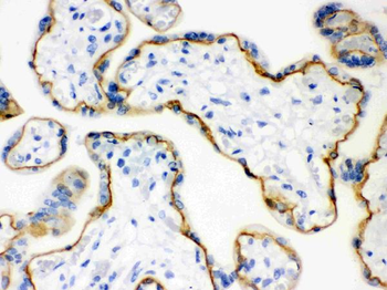
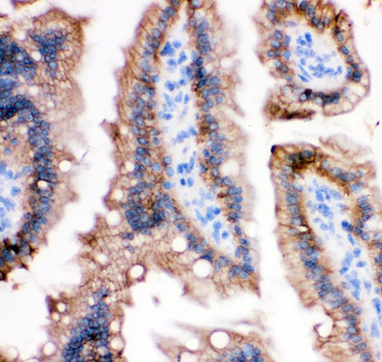
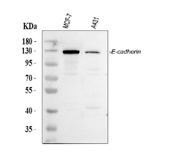
Western blot, 0.1-0.5µg/ml, Human Immunohistochemistry (Paraffin-embedded Section), 2-5µg/ml, Human, Mouse, Rat Immunocytochemistry/Immunofluorescence, 5 µg/ml, Human ELISA, 0.1-0.5µg/ml, -
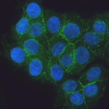
IF analysis of E Cadherin using anti-E Cadherin antibody. E Cadherin was detected in an immunocytochemical section of MCF-7 cells. Enzyme antigen retrieval was performed using IHC enzyme antigen retrieval reagent for 15 mins. The cells were blocked with 10% goat serum. And then incubated with 5 µg/mL rabbit anti-E Cadherin Antibody overnight at 4C. DyLight488 Conjugated Goat Anti-Rabbit IgG was used as secondary antibody at 1:100 dilution and incubated for 30 minutes at 37C. The section was counterstained with DAPI. Visualize using a fluorescence microscope and filter sets appropriate for the label used.
IF analysis of E Cadherin using anti-E Cadherin antibody. E Cadherin was detected in paraffin-embedded section of human colon cancer tissue. Heat mediated antigen retrieval was performed in citrate buffer (pH6, epitope retrieval solution) for 20 mins. The tissue section was blocked with 10% goat serum. The tissue section was then incubated with 5 µg/mL rabbit anti-E Cadherin Antibody overnight at 4C.DyLight488 Conjugated Goat Anti-Rabbit IgG was used as secondary antibody at 1:100 dilution and incubated for 30 minutes at 37C. The section was counterstained with DAPI. Visualize using a fluorescence microscope and filter sets appropriate for the label used.
IF analysis of E Cadherin using anti-E Cadherin antibody. E Cadherin was detected in paraffin-embedded section of human colon organoid tissue. Heat mediated antigen retrieval was performed in citrate buffer (pH6, epitope retrieval solution) for 20 mins. The tissue section was blocked with 10% goat serum. The tissue section was then incubated with 5 µg/mL rabbit anti-E Cadherin Antibody overnight at 4C. Cy3 Conjugated Goat Anti-Rabbit IgG was used as secondary antibody at 1:100 dilution and incubated for 30 minutes at 37C. The section was counterstained with DAPI. Visualize using a fluorescence microscope and filter sets appropriate for the label used.
IF analysis of E Cadherin using anti-E Cadherin antibody. E Cadherin was detected in paraffin-embedded section of human ileum tissue. Heat mediated antigen retrieval was performed in citrate buffer (pH6, epitope retrieval solution) for 20 mins. The tissue section was blocked with 10% goat serum. The tissue section was then incubated with 5 µg/mL rabbit anti-E Cadherin Antibody overnight at 4C. Cy3 Conjugated Goat Anti-Rabbit IgG was used as secondary antibody at 1:100 dilution and incubated for 30 minutes at 37C. The section was counterstained with DAPI. Visualize using a fluorescence microscope and filter sets appropriate for the label used.
IF analysis of E Cadherin using anti-E Cadherin antibody. E Cadherin was detected in paraffin-embedded section of mouse colon tissue. Heat mediated antigen retrieval was performed in citrate buffer (pH6, epitope retrieval solution) for 20 mins. The tissue section was blocked with 10% goat serum. The tissue section was then incubated with 5 µg/mL rabbit anti-E Cadherin Antibody overnight at 4C.DyLight488 Conjugated Goat Anti-Rabbit IgG was used as secondary antibody at 1:100 dilution and incubated for 30 minutes at 37C. The section was counterstained with DAPI. Visualize using a fluorescence microscope and filter sets appropriate for the label used.
IF analysis of E Cadherin using anti-E Cadherin antibody. E Cadherin was detected in paraffin-embedded section of mouse ileum organoid tissue. Heat mediated antigen retrieval was performed in citrate buffer (pH6, epitope retrieval solution) for 20 mins. The tissue section was blocked with 10% goat serum. The tissue section was then incubated with 5 µg/mL rabbit anti-E Cadherin Antibody overnight at 4C. Cy3 Conjugated Goat Anti-Rabbit IgG was used as secondary antibody at 1:100 dilution and incubated for 30 minutes at 37C. The section was counterstained with DAPI. Visualize using a fluorescence microscope and filter sets appropriate for the label used.
IF analysis of E Cadherin using anti-E Cadherin antibody. E Cadherin was detecte
* VAT and and shipping costs not included. Errors and price changes excepted