E. coli-derived rat Lipocalin 2 recombinant protein (Position: Q21-N198). Rat Lipocalin 2 shares 64.4 % and 81.1 % amino acid (aa) sequence identity with human and mouse Lipocalin 2, respectively.
Application Notes: Western blot, 0.1-0.5µg/ml, Mouse Immunohistochemistry (Paraffin-embedded Section), 0.5-1µg/ml, Mouse, Rat Immunohistochemistry (Frozen Section), 0.5-1µg/ml, Mouse, Rat ELISA (cap) ,1-5µg/ml, Rat. Add 0.2ml of distilled water will yield a concentration of 500ug/ml
IHC analysis of Lipocalin 2 using anti-Lipocalin 2 antibody. Lipocalin 2 was detected in frozen section of mouse lung tissue. The tissue section was blocked with 10% goat serum. The tissue section was then incubated with 1 µg/ml rabbit anti-Lipocalin 2 Antibody overnight at 4C. Biotinylated goat anti-rabbit IgG was used as secondary antibody and incubated for 30 minutes at 37C. The tissue section was developed using Strepavidin-Biotin-Complex (SABC) with DAB as the chromogen.
WB analysis of Lipocalin 2 using anti-Lipocalin 2 antibody.La
IHC analysis of Lipocalin 2 using anti-Lipocalin 2 antibody. Lipocalin 2 was detected in frozen section of mouse spleen tissue. The tissue section was blocked with 10% goat serum. The tissue section was then incubated with 1 µg/ml rabbit anti-Lipocalin 2 Antibody overnight at 4C. Biotinylated goat anti-rabbit IgG was used as secondary antibody and incubated for 30 minutes at 37C. The tissue section was developed using Strepavidin-Biotin-Complex (SABC) with DAB as the chromogen.
IHC analysis of Lipocalin 2 using anti-Lipocalin 2 antibody. Lipocalin 2 was detected in frozen section of rat lung tissue. The tissue section was blocked with 10% goat serum. The tissue section was then incubated with 1 µg/ml rabbit anti-Lipocalin 2 Antibody overnight at 4C. Biotinylated goat anti-rabbit IgG was used as secondary antibody and incubated for 30 minutes at 37C. The tissue section was developed using Strepavidin-Biotin-Complex (SABC) with DAB as the chromogen.
IHC analysis of Lipocalin 2 using anti-Lipocalin 2 antibody. Lipocalin 2 was detected in frozen section of rat spleen tissue. The tissue section was blocked with 10% goat serum. The tissue section was then incubated with 1 µg/ml rabbit anti-Lipocalin 2 Antibody overnight at 4C. Biotinylated goat anti-rabbit IgG was used as secondary antibody and incubated for 30 minutes at 37C. The tissue section was developed using Strepavidin-Biotin-Complex (SABC) with DAB as the chromogen.
IHC analysis of Lipocalin 2 using anti-Lipocalin 2 antibody. Lipocalin 2 was detected in paraffin-embedded section of Mouse Spleen Tissue. Heat mediated antigen retrieval was performed in citrate buffer (pH6, epitope retrieval solution) for 20 mins. The tissue section was blocked with 10% goat serum. The tissue section was then incubated with 1 µg/ml rabbit anti-Lipocalin 2 Antibody overnight at 4C. Biotinylated goat anti-rabbit IgG was used as secondary antibody and incubated for 30 minutes at 37C. The tissue section was developed using Strepavidin-Biotin-Complex (SABC) with DAB as the chromogen.
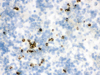
IHC analysis of Lipocalin 2 using anti-Lipocalin 2 antibody. Lipocalin 2 was detected in paraffin-embedded section of Rat Spleen Tissue. Heat mediated antigen retrieval was performed in citrate buffer (pH6, epitope retrieval solution) for 20 mins. The tissue section was blocked with 10% goat serum. The tissue section was then incubated with 1 µg/ml rabbit anti-Lipocalin 2 Antibody overnight at 4C. Biotinylated goat anti-rabbit IgG was used as secondary antibody and incubated for 30 minutes at 37C. The tissue section was developed using Strepavidin-Biotin-Complex (SABC) with DAB as the chromogen.
Western blot analysis of Lipocalin 2 using anti-Lipocalin 2 antibody. Electrophoresis was performed on a 5-20% SDS-PAGE gel at 70V (Stacking gel) / 90V (Resolving gel) for 2-3 hours. The sample well of each lane was loaded with 50 ug of sample under reducing conditions. Lane 1: Mouse Lung Tissue Lysate, Lane 2: Mouse Intestine Tissue Lysate. After Electrophoresis, proteins were transferred to a Nitrocellulose membrane at 150mA for 50-90 minutes. Blocked the membrane with 5% Non-fat Milk/ TBS for 1.5 hour at RT. The membrane was incubated with rabbit anti-Lipocalin 2 antigen affinity purified polyclonal antibody at 0.5 µg/mL overnight at 4C, then washed with TBS-0.1% Tween 3 times with 5 minutes each and probed with a goat anti-rabbit IgG-HRP secondary antibody at a dilution of 1:10000 for 1.5 hour at RT. The signal is developed using an Enhan
* VAT and and shipping costs not included. Errors and price changes excepted