
ACTA2 antibody, Unconjugated, Rabbit, Polyclonal
Catalog Number:
BYT-ORB323289-IH
- Images (9)


| Article Name: | ACTA2 antibody, Unconjugated, Rabbit, Polyclonal |
| Biozol Catalog Number: | BYT-ORB323289-IH |
| Supplier Catalog Number: | orb323289 |
| Alternative Catalog Number: | BYT-ORB323289-100,BYT-ORB323289-500 |
| Manufacturer: | Biorbyt |
| Host: | Rabbit |
| Category: | Antikörper |
| Application: | ELISA, IHC-P, WB |
| Species Reactivity: | Human, Mouse, Rat |
| Immunogen: | KLH conjugated synthetic peptide derived from human ACTA2. Please contact us for the exact immunogen sequence. The peptide is available as orb374950. |
| Conjugation: | Unconjugated |
| Alternative Names: | Anti-a actin antibody, anti-AAT6 antibody, anti-ACTA_HUMAN antibody, anti-ACTA2 antibody, anti-Actin alpha 2 smooth muscle aorta antibody, anti-Actin aortic smooth muscle antibody, anti-Actin, aortic smooth muscle antibody, anti-ACTSA antibody, anti-ACTVS antibody, anti-Alpha 2 actin antibody, anti-Alpha actin 2 antibody, anti-Alpha cardiac actin antibody, anti-alpha sma antibody, anti-Alpha-actin-2 antibody |
| ACTA2 antibody |
| Application Dilute: | WB: 1:100-2000, IHC-P: 1:100-1000 (based on 0.5 mg/ml) |
|
|
Immunofluorescence analysis of mouse uterus tissue using anti-ACTA2 (dilution of primary antibody - 2.5 ug/ml) |
|
|
IF image of mouse uterus tissue using ACTA2 antibody (primary antibody at 2.5 ug/ml) |
|
|
Immunohistochemical staining of paraffin embedded mouse brain tissue using ACTA2 antibody (primary antibody at 2.5 ug/ml) |
|
|
IHC-P image of mouse heart tissue using ACTA2 antibody (dilution of primary antibody at 2.5 ug/ml) |
|
|
IHC-P staining of mouse uterus tissue using anti-ACTA2 (dilution at 2.5 ug/ml) |
|
|
Immunohistochemical staining of mouse uterus tissue using anti-ACTA2 (dilution of primary antibody - 2.5 ug/ml) |
|
|
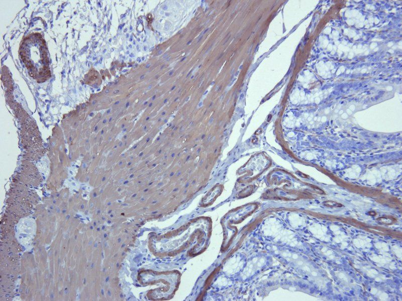
Immunohistochemical staining of paraffin embedded rat colon tissue using ACTA2 antibody (primary antibody at 2.5 ug/ml) |
|
|
IHC-P image of rat colon tissue using anti-ACTA2 (dilution of primary antibody at 2.5 ug/ml) |
|
|
Western blot analysis of rat muscle (lane 1), rat heart (lane 2), rat brain (lane 3), rat liver (lane 4), rat lung (lane 5), rat pancreas (lane 6) using ACTA2 antibody (1 ug/ml) |
Product Guarantee and Expert Support
