136.36mM Ethanolamine, 133.23 mM Chlorides, 9.55mM Phosphates, 9.55mM Sodium Bicarbonate.
Target:
Beclin 1
Application Dilute:
WB (1:1000), ICC/IF (1:100)
Application Notes:
Application Notes: A 1:1000 dilution of SPC-601 was sufficient for detection of Beclin1 on 293T lysates using Goat anti-rabbit IgG:HRP as the secondary antibody
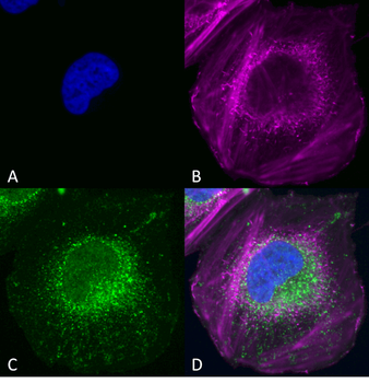
Immunocytochemistry/Immunofluorescence analysis using Rabbit Anti-Beclin 1 Polyclonal Antibody. Tissue: SK-N-BE. Species: Human. Primary Antibody: Rabbit Anti-Beclin 1 Polyclonal Antibody at 1:200 for Overnight. Secondary Antibody: Anti-Rabbit: AlexaFluor 555 at 1:1000. Counterstain: Hoechst, Phalloidin AlexaFluor 647 at 1:1000. Localization: Mitochondria, endosomes/exosomes. A) Hoechst Nuclear Stain B) Phalloidin AlexaFluor 647 C) Anti-Beclin 1 D) Merge.
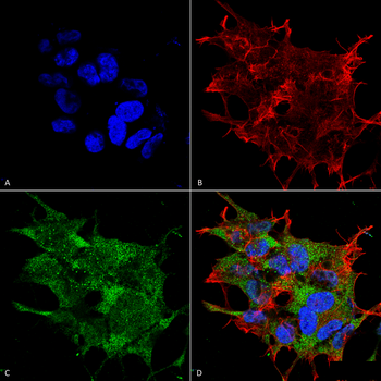
Immunocytochemistry/Immunofluorescence analysis using Rabbit Anti-Beclin 1 Polyclonal Antibody. Tissue: Neuroblastoma cell line (SK-N-BE). Species: Human. Fixation: 4% Formaldehyde for 15 min at RT. Primary Antibody: Rabbit Anti-Beclin 1 Polyclonal Antibody at 1:100 for 60 min at RT. Secondary Antibody: Goat Anti-Rabbit ATTO 488 at 1:100 for 60 min at RT. Counterstain: Phalloidin Texas Red F-Actin stain, DAPI (blue) nuclear stain at 1:1000, 1:5000 for 60min RT, 5min RT. Localization: Golgi apparatus, Dendrites and Cell bodies of cerebellar Purkinje cells. Magnification: 60X. (A) DAPI (blue) nuclear stain (B) Phalloidin Texas Red F-Actin stain (C) Beclin 1 Antibody (D) Composite.
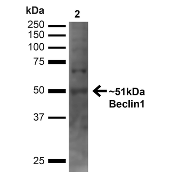
Western blot analysis of Human Cervical cancer cell line (HeLa) lysate showing detection of ~51kDa Beclin 1 protein using Rabbit Anti-Beclin 1 Polyclonal Antibody. Lane 1: MW Ladder. Lane 2: Human HeLa (20 µg). Load: 20 µg. Block: 5% milk + TBST for 1 hour at RT. Primary Antibody: Rabbit Anti-Beclin 1 Polyclonal Antibody at 1:1000 for 1 hour at RT. Secondary Antibody: Goat Anti-Rabbit: HRP at 1:2000 for 1 hour at RT. Color Development: TMB solution for 12 min at RT. Predicted/Observed Size: ~51kDa.
Western blot analysis of Human Embryonic kidney epithelial cell line (HEK293T) lysate showing detection of ~51kDa Beclin 1 protein using Rabbit Anti-Beclin 1 Polyclonal Antibody. Lane 1: MW Ladder. Lane 2: Human 293T (20 µg). Load: 20 µg. Block: 5% milk + TBST for 1 hour at RT. Primary Antibody: Rabbit Anti-Beclin 1 Polyclonal Antibody at 1:1000 for 1 hour at RT. Secondary Antibody: Goat Anti-Rabbit: HRP at 1:2000 for 1 hour at RT. Color Development: TMB solution for 12 min at RT. Predicted/Observed Size: ~51kDa.
* VAT and and shipping costs not included. Errors and price changes excepted